


Oral Cancer
Image-guided techniques for biopsy site selection and tumour boundary delineation have the potential to significantly improve the management of oral cancer. OCT can provide high-resolution 3-dimensional images of tissue in real time to distinguish morphological tissue features associated with neoplastic development.
Optical Coherence Tomography (OCT)
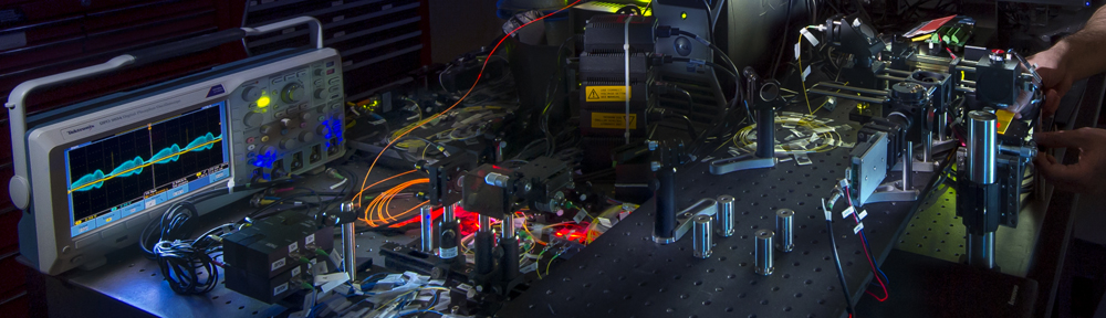
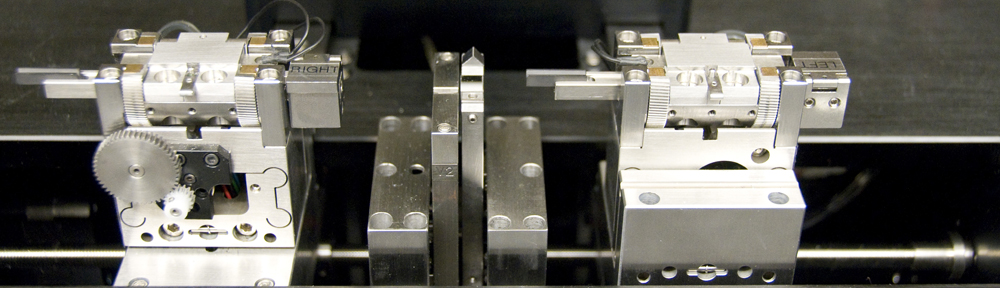
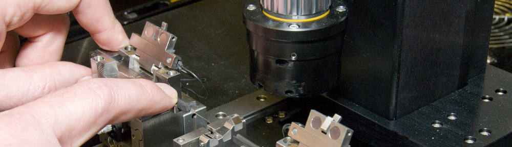
Our OCT catheter and swept-source instrumentation is capable of wide-field in vivo imaging in the oral cavity. We use a hand-held side-looking fiber-optic rotary pullback catheter that can cover two dimensional tissue imaging fields approximately 2.5 mm wide by up to 90 mm length in a single image acquisition. This instrument provides a wide-field view of features such as epithelial thickness and continuity of the basement membrane that may be useful in clinic for chair-side management of oral lesions.
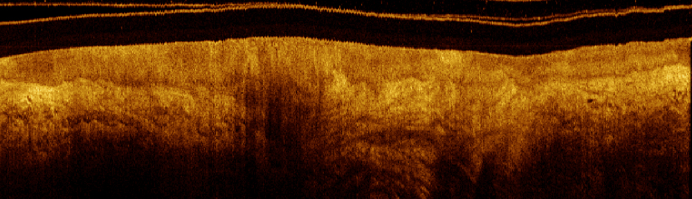
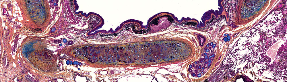
The OCT sections below show normal (top) and abnormal (bottom) oral mucosa. The images are very long (33 mm) and narrow (1.2 mm) and have been compressed in the horizontal dimension to facilitate display (28:1 aspect). The normal section shows the epithelium and submucosa as uniform intensity stratifications.
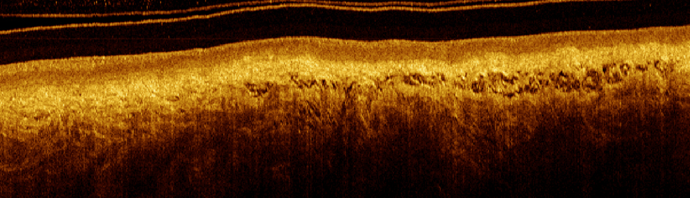
The abnormal section shows thickening of the epithelium, loss of visibility of the basement membrane, and irregular intensity stratification.