


Research
Cancer is most treatable when it is detected early. The Biophotonics Group at the BC Cancer Agency (BCCA) develops optical tools for early detection and disease management.
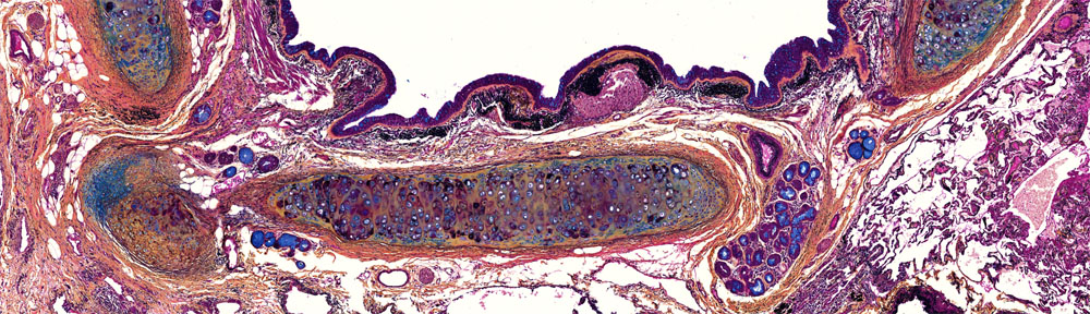
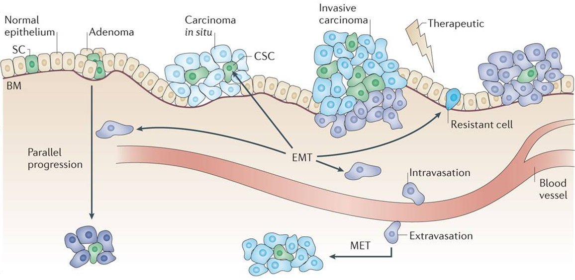
The imaging team is interested in the application of light for the early detection of epithelial cancers using in vivo optical imaging. The majority of cancers – about 80% in fact – originate in the epithelium, the thin layer of tissue that covers our body (skin) and lines our hollow organs (oral cavity, GI tract, lungs, for example). Carcinomas – cancers that originate in the epithelium – may spread to other parts of the body when one of the cancer cell passes through the basement membrane (invasion), penetrates a vessel of the circulatory system, and some time later, re-penetrates the vessel to form a secondary tumor (MET or metastasis).
While traditional imaging modalities such as computed tomography (CT) and magnetic resonance imaging (MRI) are able to image these organs from outside the body, the resolution is insufficient to detect cancer an its earliest stages.
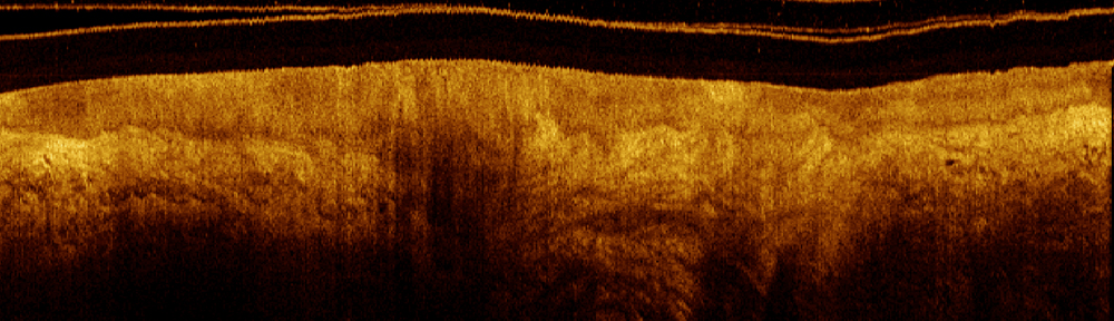
Optical imaging on the other hand has very high resolution but lacks the penetration depth to image organs from outside the body. Small endoscope-compatible imaging tools, however, can access these sites to provide detailed imagery at very high resolution. In fact, the penetration depth of optical coherence tomography (OCT) – one of the technologies of interest to our group – is sufficient to image the entire epithelial thickness and sub-epithelial structure and function in the stroma below.
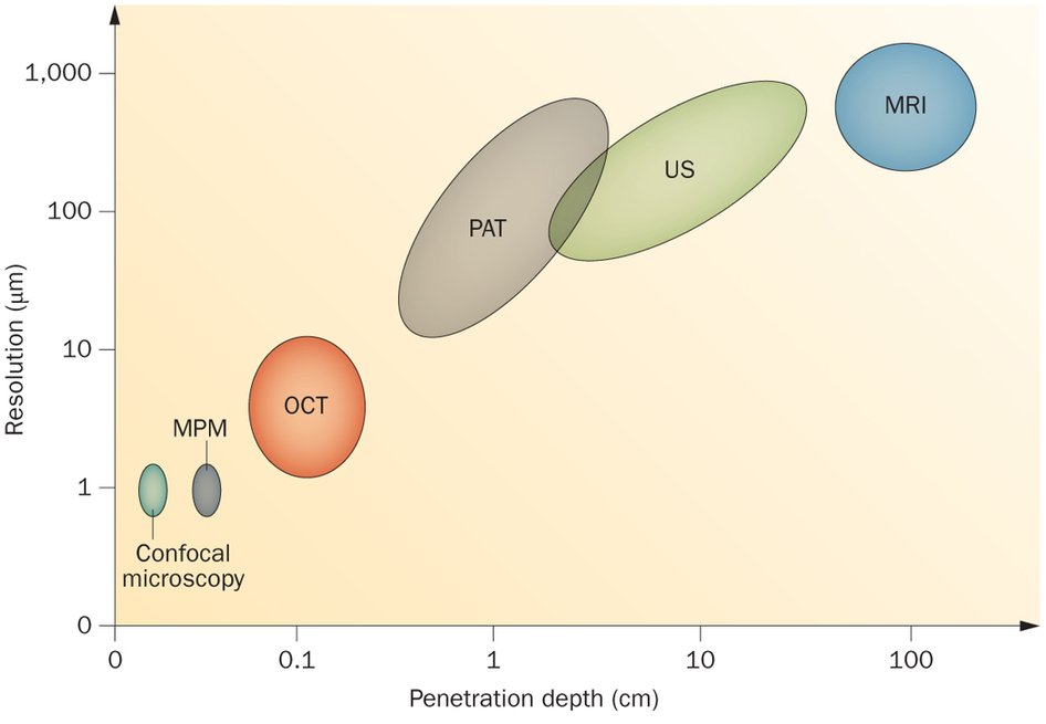
The figure below illustrates the resolution and penetration depth of the ultrasound (US) and magnetic resonance imaging (MRI) with some up-and-coming research imaging modalities including optical coherence tomography (OCT), photoacoustic tomography (PAT), multiphoton microscopy (MPM) and confocal microscopy.
Some of the earliest changes due to a pre-cancer cancerous lesion are detectable in the epithelium and underlying stroma. Optical imaging using reflectance and fluorescence contrast can identify the small structural and functional biomarkers that are associated with cancer progression, often enabling detection before the lesion actually becomes a cancer.
The pages linked below describe some of the in vivo optical imaging technologies we are developing and some of the clinical applications where they add value to detection, diagnosis and management of cancer.