

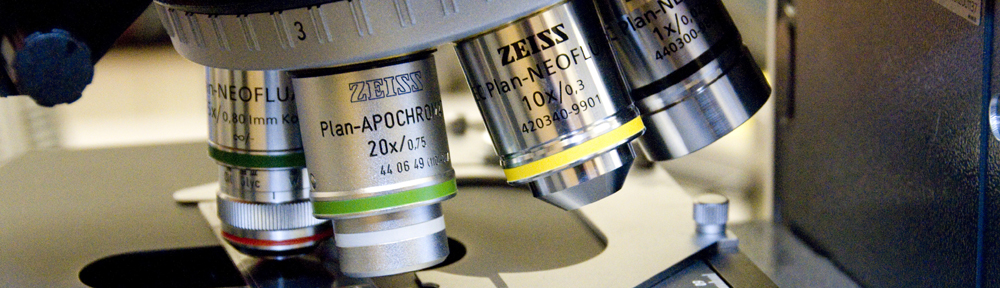


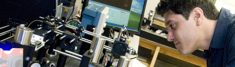


Confocal microscopy is used in the laboratory to study the 3-D structure and function of tissue and cells at high resolution. Our group develops miniature clinic-ready confocal instruments to acquire real-time confocal images of tissue structure and function in vivo for the detection and management of cancer. Some of the confocal devices are small enough for deployment through an endoscope.
Confocal microscopy provides high-resolution thin optical sections from just below the tissue surface. The axial and lateral resolution is determined by the wavelength and numerical aperture of the system. Reflectance and fluorescent contrast is possible, however, the majority of the instruments we develop use confocal florescence to maximize signal-to-noise ratio.
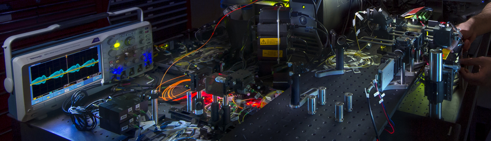
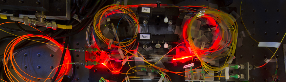
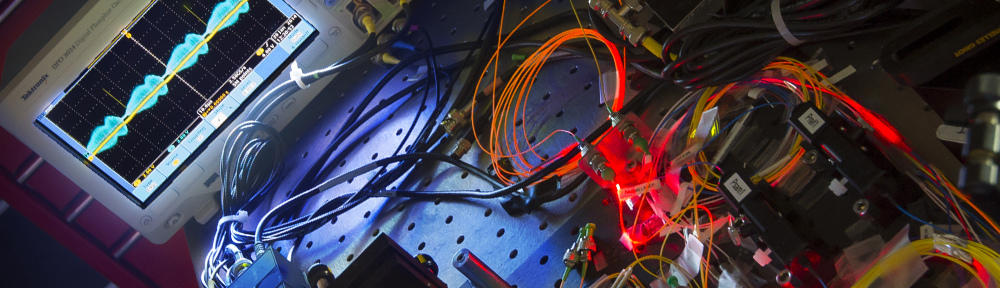
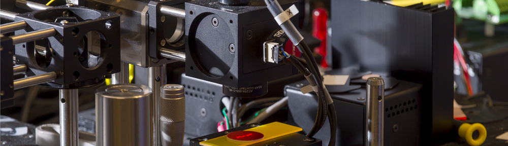
Instrumentation
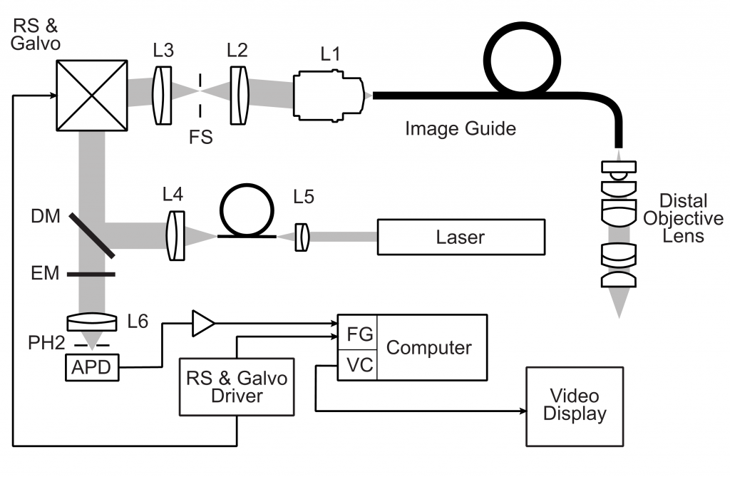
Our system employs a fiber-optic image guide with 30,000 fibers and distal scanning by a galvanometer and resonance scanner. Excitation light is provided by a 445 nm laser diode. Emission passed by 84 nm bandpass filter centered at 510 nm is detected by an avalanche photo diode (APD). The custom 7-element objective lens (3X/1.0NA) is corrected for axial chromatic aberration. Lateral (1.4 µm) and axial resolution (10 µm) enables the visualization of cell nuclei with optical sectioning at depths up to 50µm (dye-penetration limited). The field of view is 240 µm and the frame rate is 7 Hz.
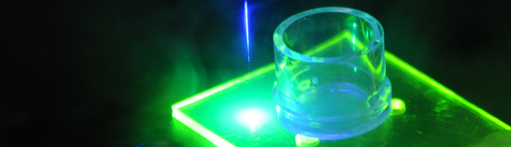
Contrast Agents
We use topically applied vital fluorescent dyes that have already been clinically approved. In a recent study we tested five dyes in vitro on cultured human cells and validated performance ex vivo on human oral mucosa. Acriflavine hydrochloride provided the best cellular contrast by preferentially staining the nuclei of the epithelium. The maximum imaging depth is limited to about 50 µm by the diffusion of the topically applied stain.
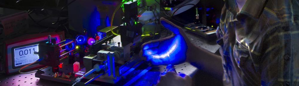
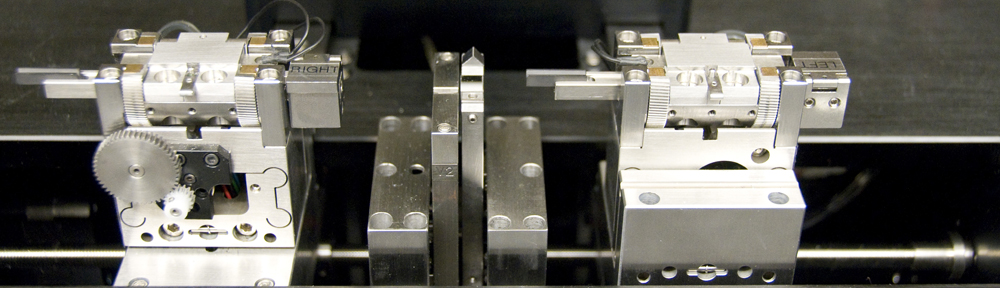
Handheld Confocal Wands
Two handheld confocal wands –one forward looking and one side-looking – were designed to accommodate the different imaging geometries of the cervix and oral cavity. The forward-looking cervical wand is used between the blades of a speculum for head-on imaging of the cervix.
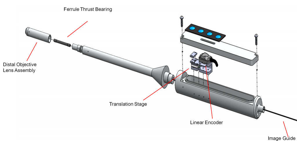
The oral wand has a fold mirror between the main lens groups of the objective lens to provide a side-looking geometry for imaging the oral cavity. In both designs, depth scanning is implemented by translating the image guide along its axis using a piezo linear stage. An encoder enables the imaging depth to be recorded.
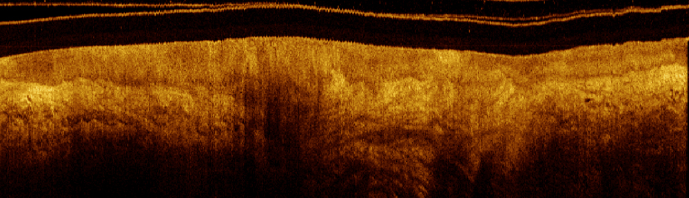
Image Analysis and Tissue Discrimination
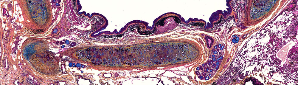
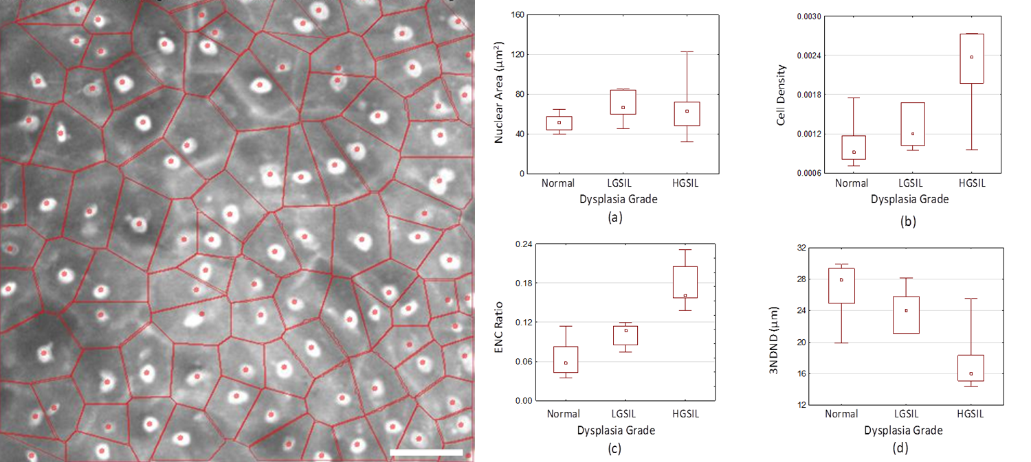
Visualization of cell nuclei enables the evaluation of cellular-level tissue morphology and architecture. The position of nuclei determined manually (or automatically) from the confocal sections can be used to calculate morphometric biomarkers of cancer such as cellular density and nuclear size. These features, measured as a function of depth from the tissue surface, can be used to discriminate normal tissue from that which is likely to progress to cancer (pre-cancer).
Continue with Clinical Applications.