


Cervical cancer remains a tragic cause of mortality in Canada, the United States (US), and the rest of the developed world. It is particularly devastating in low and middle-income countries (LMICs) where 90% of all deaths due to cervical cancer occur. Despite this expensive screening infrastructure, invasive cancers still occur and consequently there remains an unmet clinical need for better screening and diagnostic techniques.
Confocal Fluorescence
Our handheld confocal wand allows the collection of optical sections from just below the surface of the cervix. Topically applied Acriflavine Hydrochloride provides nuclear contrast. The movie below shows a uniform field of cells suggesting normal cervical mucosa.
Mutispectral Imaging
Devices for fluorescence and reflectance imaging (“Multispectral Imaging”) use a large field of view to image the entire cervix and provide multispectral images with both fluorescence and reflectance contrast. Our team has built and evaluated a device for multispectral imaging called the multispectral digital colposcope (MDC3). As a regular colposcope, the MDC3 illuminates a 3cm diameter area of the cervix with white light at 3X magnification. Additionally, the MDC3 provides three fluorescence images with UV (350-360 nm), violet (410-430 nm), and blue (450-460 nm) excitation, and two reflectance images with violet and blue illumination (6 images total).
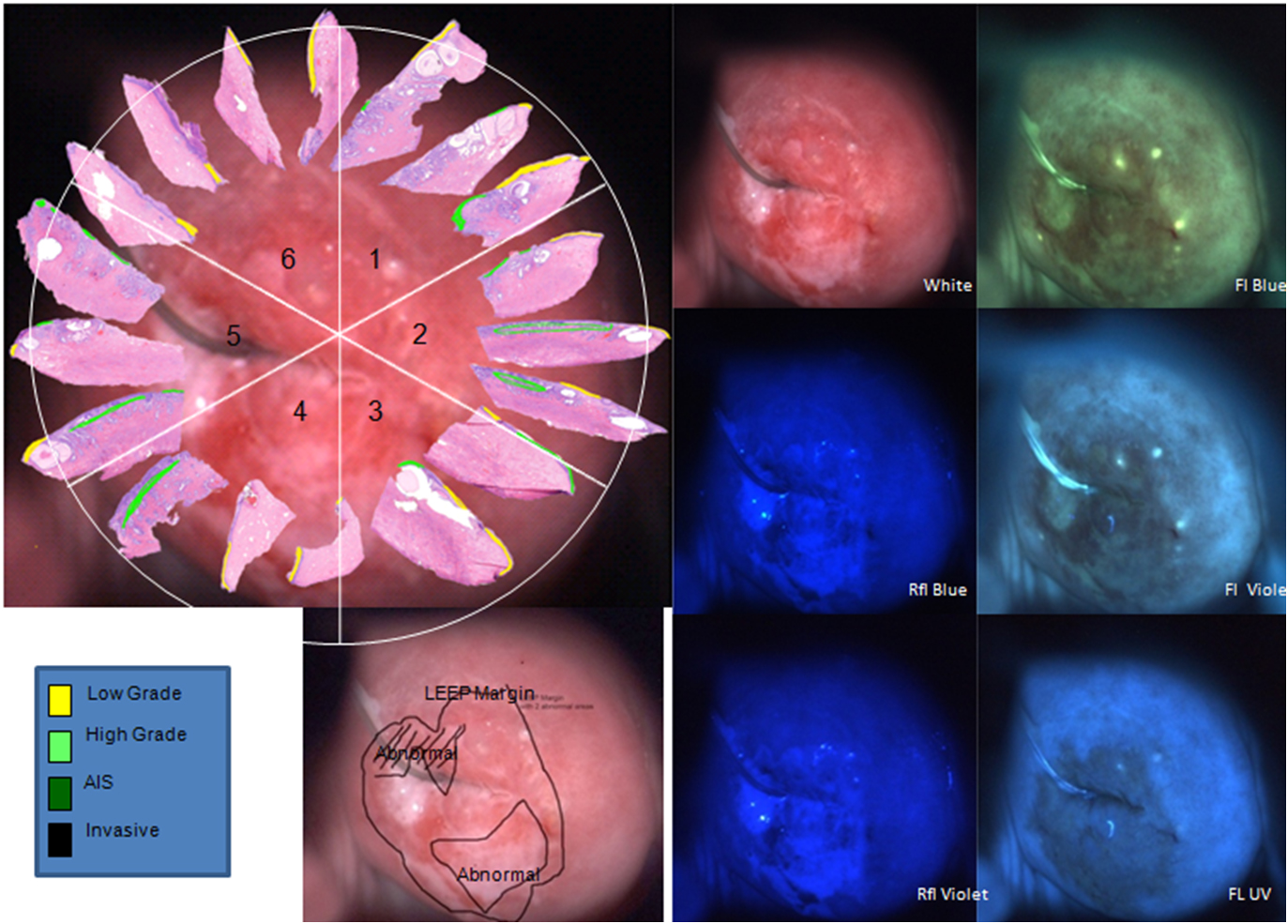
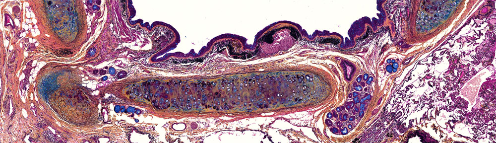
The figure shows reflectance and fluorescence imaging of the cervix by the MDC3. Top left panel shows histological sections annotated with diagnosis for the six sextants. The multispectral images from the MDC are shown on the right as a 2×3 array of images with reflectance and fluorescence in the first and second columns, respectively. The clinical impression of the colposcopist is annotated on the white-light images on the bottom left.