Rising Stars in Integrative Oncology

Dr. Adrian Tanskanen in the MacAulay lab has been selected as a BC Cancer Rising Star (post-doctoral award).

The Rising Stars program was established to celebrate Dr. Connie Eaves’ Gairdner Wightman Award, her election to the Royal Society in the UK and her appointment to the Order of Canada. A pioneer in her field, Dr. Eaves has been a mentor to many young researchers at BC Cancer and a long-time advocate for equal opportunities in cancer research.

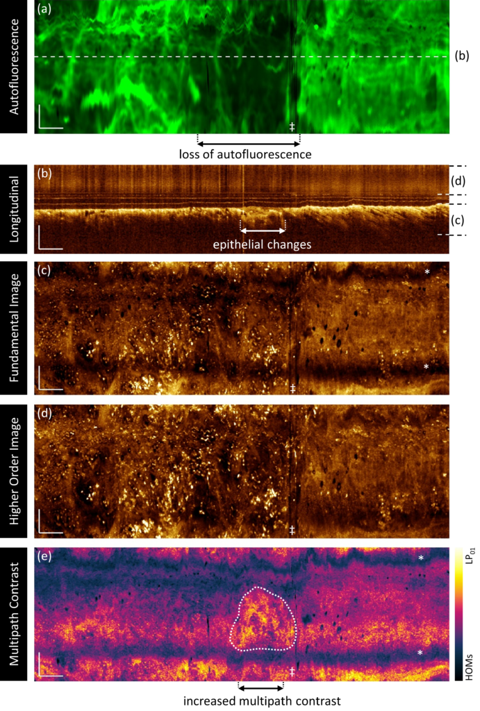
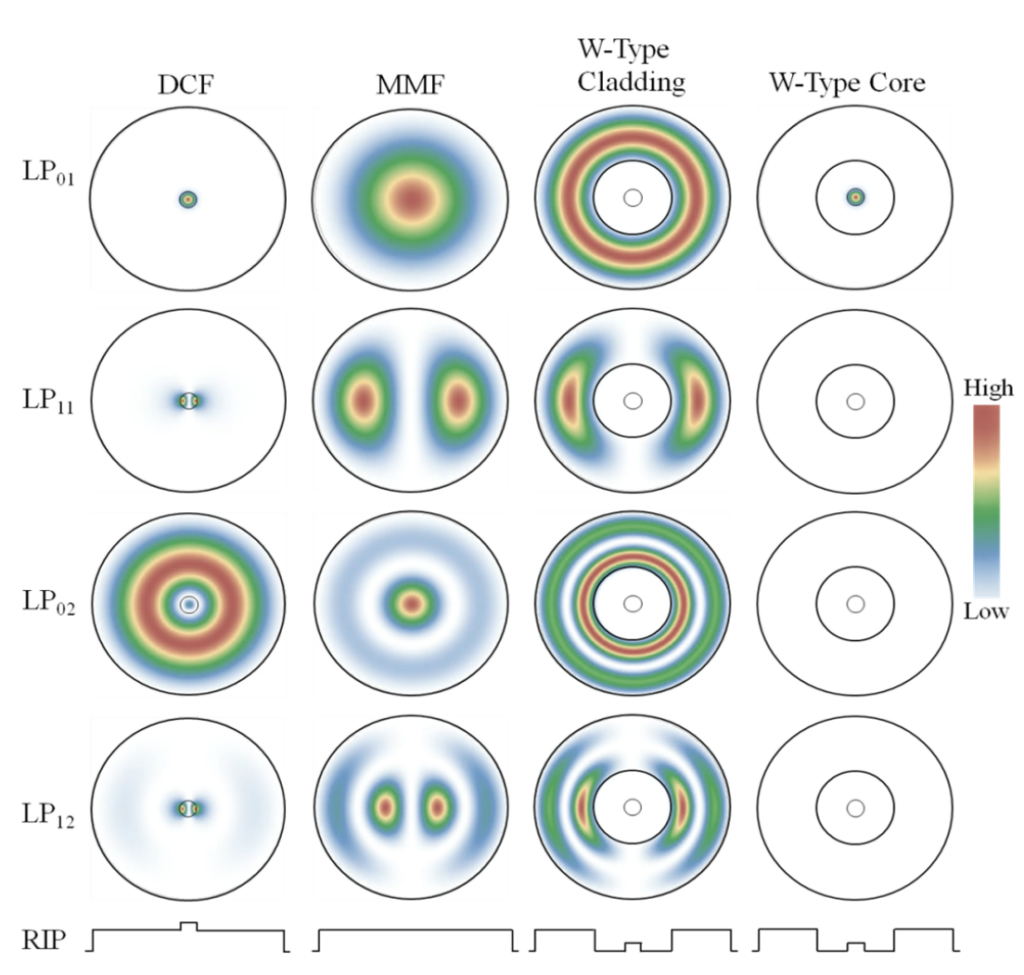
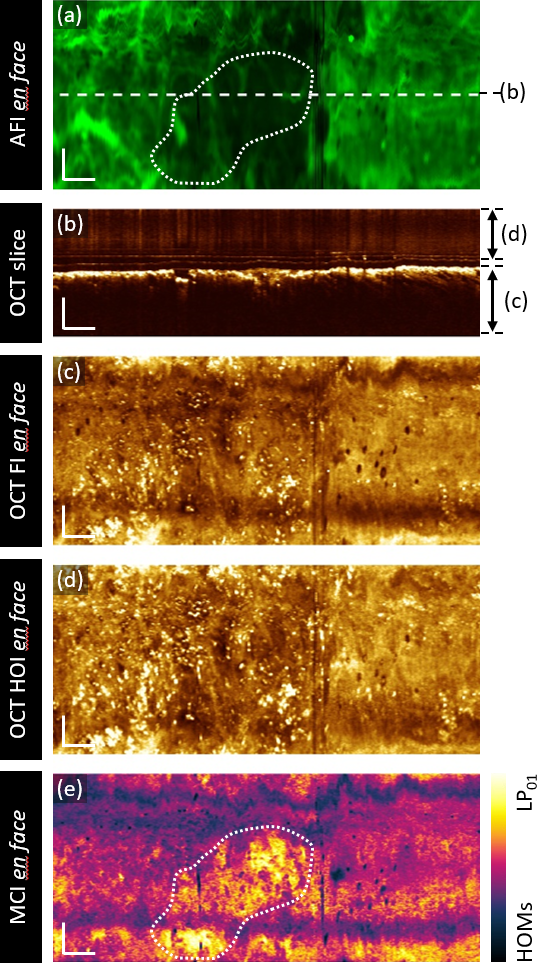
Dr. Tanskanen’s research focuses on developing angular diversity contrast mechanisms in novel optical endoscopes for lung cancer detection. This builds off of a recent publication and patent which demonstrated potential for using higher-order modes in optical fibers to collect multiple simultaneous images of a sample.

New publication on machine learning tools for lung cancer management

Ian Janzen, a PhD student in our group has a new publication in MDPI Cancers.

Lung cancer is the leading cause of cancer deaths in Canada. Most lung cancers are diagnosed at advanced stages, where treatment pathways are determined by target mutations. Current standard of care for patients with cancers with a high PD-L1 score is an immunotherapy regimen – however, nearly half of patients do not respond to this approach.

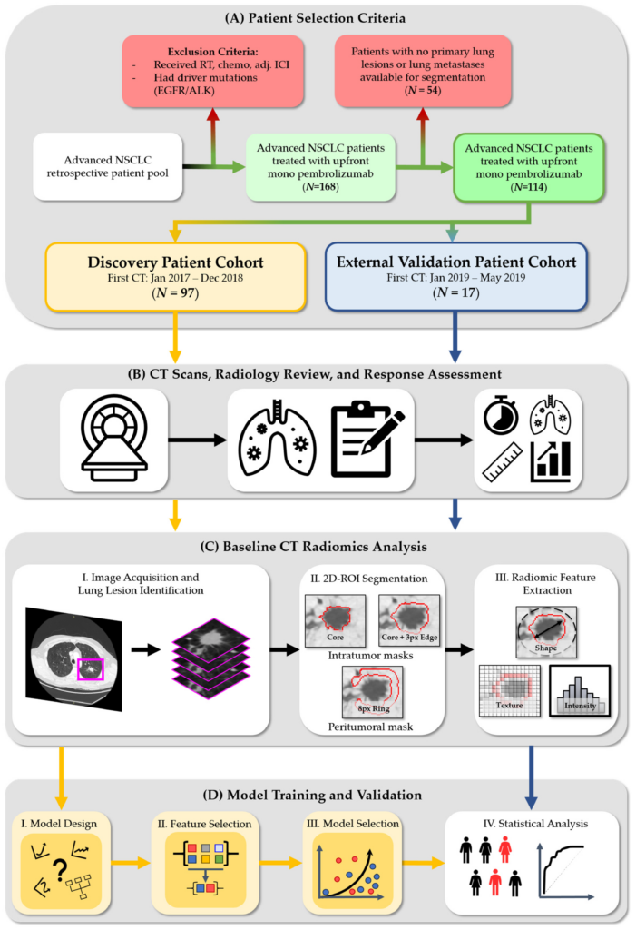
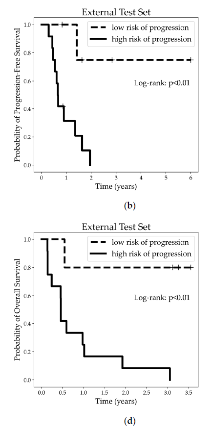
Ian’s work develops a predictive risk model using a combination of imaging features (‘radiomics’) derived from CT scans before treatment and clinical descriptors to determine which patient cohorts will not benefit from standard immunotherapy treatments.

Congratulations Dr. Malone!

Congratulations to Dr. Jeanie Malone for successfully defending her PhD thesis!

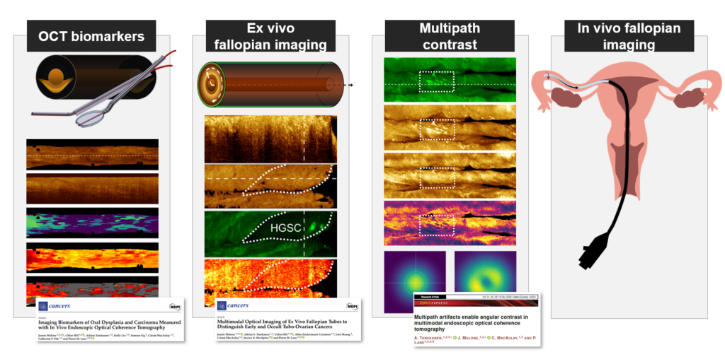
Jeanie’s work ‘Towards endoscopic optical imaging of the fallopian tubes for tubo-ovarian cancer detection explored the use of optical imaging catheters for cancer detection and management. Her work focused on detecting the earliest ovarian cancers where they form in the fallopian tubes. She demonstrated these devices can distinguish disease from normal tissue – the first step towards developing screening tools for ovarian cancer detection.

Select Publications:

New publication on OCT-AFI biomarkers for tubo-ovarian cancer detection

Sample imaging of a high-grade serous ovarian carcinoma.

Jeanie, a PhD student in our lab has a new publication in MDPI Cancers.

Tubo-ovarian cancers are associated with high mortality. Early diagnosis is associated with better patient outcomes, but there are currently no effective screening measures. The most common and aggressive ovarian cancers originate in the ends of the fallopian tubes.

This paper explores whether a previously developed optical imaging catheter can detect early or occult lesions in the fallopian tubes. This device collects three-dimensional structural images of tissue through optical coherence tomography (OCT) simultaneously with functional imaging through autofluorescence imaging (AFI).

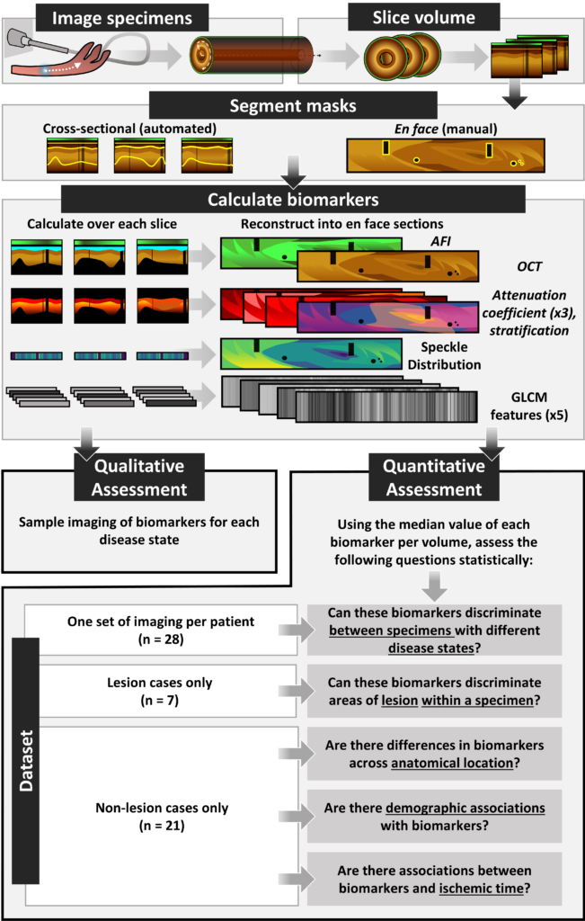
In this study, we imaged ex vivo fallopian tubes from and explored eleven imaging biomarkers for their ability to distinguish early or otherwise undetectable disease.

We found that cancers can be visually distinguished through this approach, and that there are measurable changes within the area of lesion and throughout the specimen.

This approach shows promise and merits further investigation of its diagnostic potential.

Congratulations Dr. Tanskanen!

Congratulations to Dr. Adrian Tanskanen who successfully defended his PhD thesis!

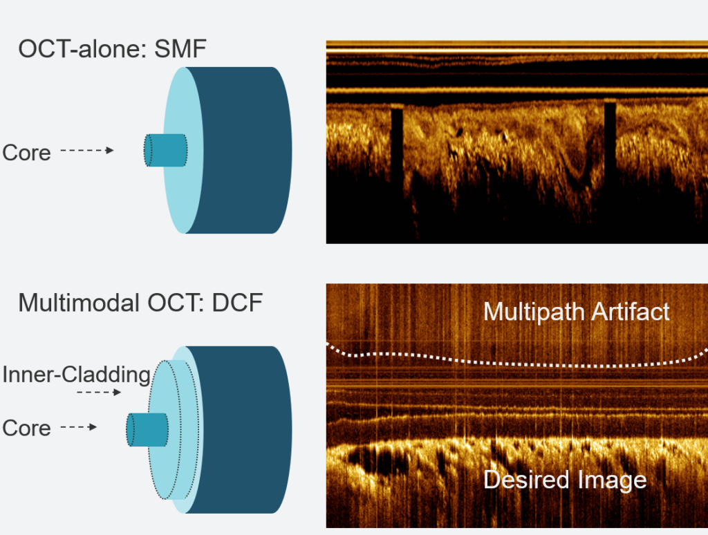
Adrian’s work ‘Leveraging Multipath Effects in Multimodal Optical Coherence Tomography for Cancer Detection‘ described methods to characterize, mitigate and leverage multipath artifacts inherent to OCT systems using double-clad fiber.

Select Publications:

New publication on OCT biomarkers of oral dysplasia and carcinoma

Jeanie, a PhD student in our lab has a new publication in MDPI Cancers.

Oral cancers are associated with high mortality in advanced stages. Early diagnosis is associated with better patient outcomes, but this is challenging to achieve as benign lesions look similar to lesions of concern, and multiple biopsies may be required to ensure the most pathologic tissue is sampled. 

Optical coherence tomography is a noninvasive imaging technique that provides three-dimensional visualization of subsurface tissue structures. We have previously developed an OCT endoscope which can reach most sites in the oral cavity – but assessing large three-dimensional volumes for the features most relevant to oral cancer is challenging.

This work leverages the work of Chloe, a recent Masters student in our lab, who developed a deep learning segmentation tool to rapidly detect the tissue surface and bottom of the epithelium in oral OCT. From these segmentations, we measure seven imaging biomarkers and assess their utility in distinguishing oral pre-cancers and cancers.

Sample imaging of severe dysplasia

New publication on endoscopic oral OCT segmentation with deep learning

Congratulations to Chloe, a recent Masters student in our lab for her first publication in MDPI Cancers.

OCT produces a large amount of 3-dimensional data which is hard to quantify manually. This study presents a neural network pipeline to simplify OCT interpretation by providing information about epithelial depth and stratification through simple en face maps. The pipeline’s predictions demonstrate as-good-as or better agreement than inter-rater agreement, suggesting strong predictive power.

Grade 2 dysplasia of the lateral tongue, with an epithelial depth heatmap in (b).
Contralateral image of the lateral tongue, with an epithelial depth heatmap in (b).

Double-Clad Fiber article in BioPhotonics

Castor Optics has published a new article on multimodal optical coherence tomography using double-clad fiber in BioPhotonics, citing one of our previous publications. Multimodal OCT often uses double-clad fiber couplers from Castor Optics.

In vivo OCT and autofluorescence imaging (AFI) of human peripheral airways using a double-clad fiber (DCF) coupler. AFI of an airway (a), magnified AFI region (b), and OCT cross sections corresponding to the dashed lines (c-e). 

SPIE Photonics West 2024

Left to right: Eric, Kimiya, Jeanie, Alicia, and Adrian

OCIL trainees presented work at SPIE Photonics West 2024 in San Francisco, California. You can read more about their presentations here:

Congratulations Mehar & Allan!

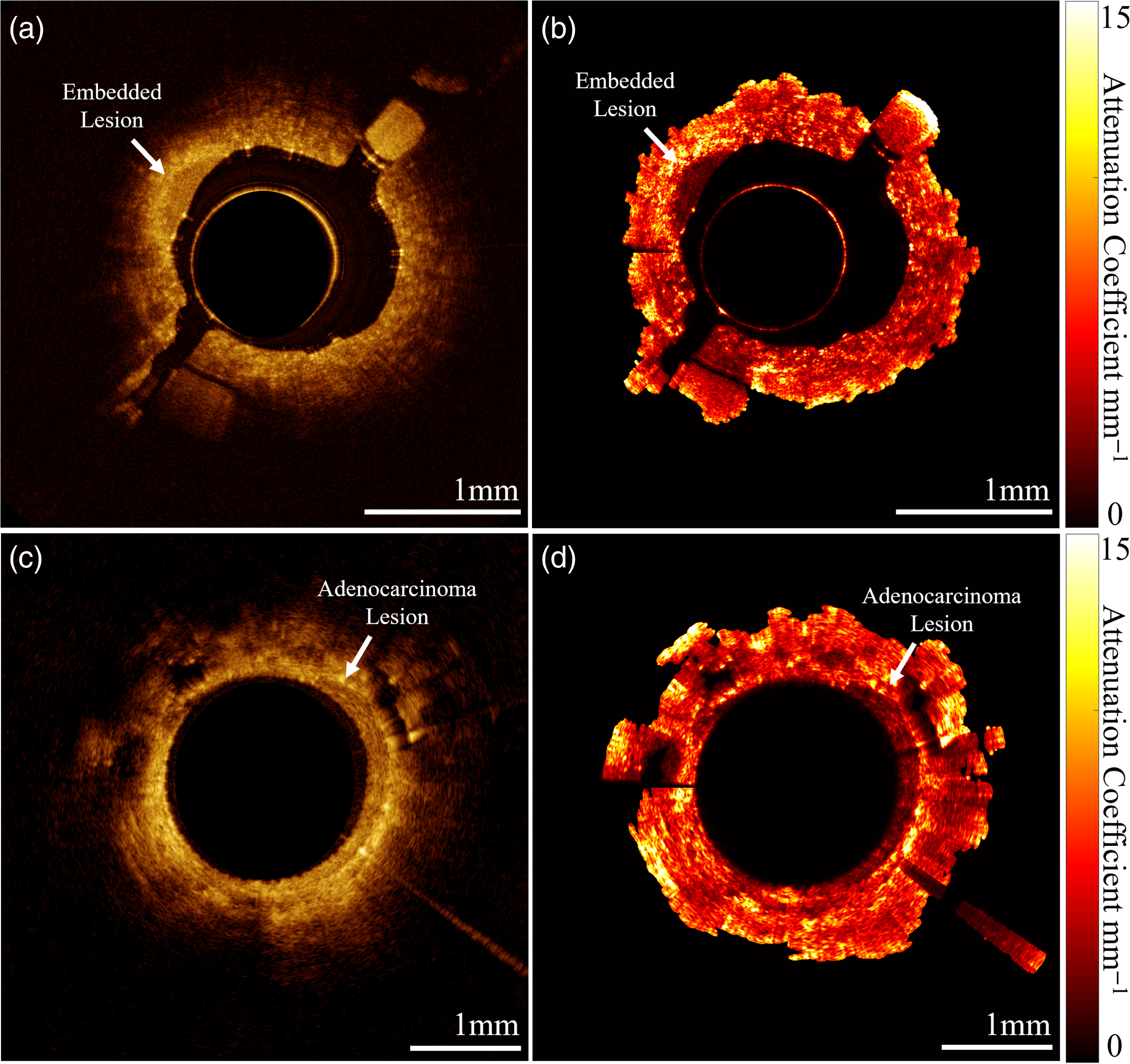
Attenuation coefficient of normal (left) and pathologic (right) oral tissue.

A segmented airway and example cross-sections (segmentations in green).

Congratulations to Mehar and Allan who successfully defended their undergraduate honours theses this fall!

Mehar’s work ‘Methods for the Estimation of Depth-resolved Attenuation in OCT‘ compared different approaches for calculating the attenuation coefficient from OCT data. This allows us to characterize optical properties of tissue as they change with respect to depth.

Allan developed an automated tool to segment the tissue surface in OCT of the small airways of the lung. This tool allow us to quantify image features in the small airways without requiring manual segmentation. You can read his thesis, ‘Lumen Segmentation in Endobronchial Optical Coherence Tomography with Deep Learning‘ online.