


Eric Brace, a PhD student in our group has a new publication in SPIE’s Journal of Biomedical Optics.
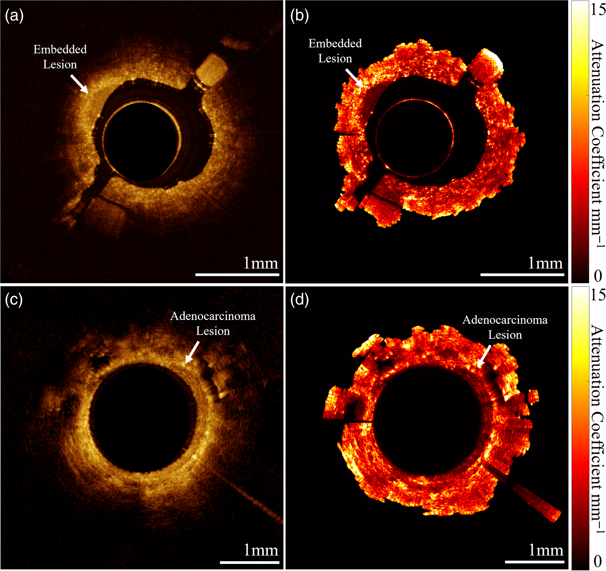
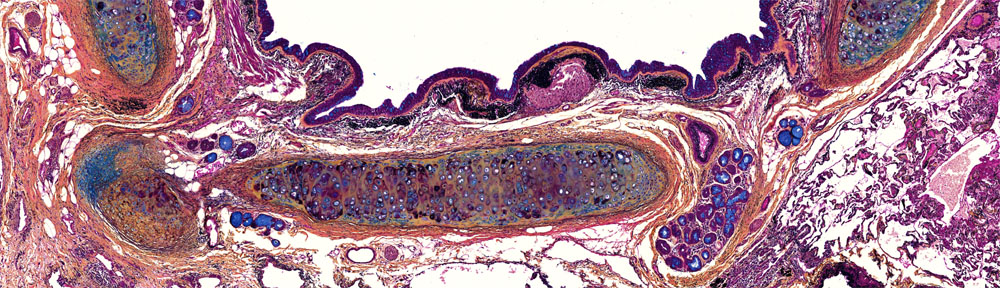
Developing new imaging devices is challenging – you might not always be able to get sample tissue to test their performance. Optical tissue ‘phantoms’ are artificial duplicates which mimic the way light interacts with tissue, acting as a test-bench for early device prototyping.
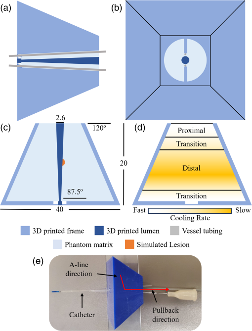
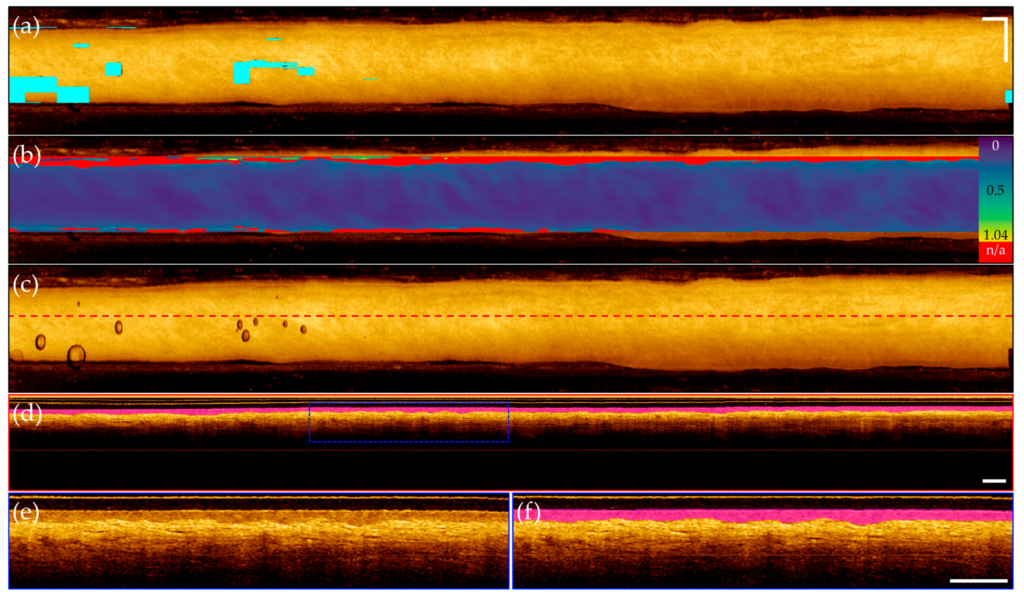
Eric developed methods to produce easy to fabricate, repeatable, 3D phantoms which mimic the small airways of the lung and surrounding parenchyma. He tailored the optical properties of these phantoms to work at the near-infrared wavelengths of our OCT systems (1310 ± 50nm). This is an important consideration as many phantoms in literature are produced for applications using visible light.
The phantoms are produced filling a 3D printed mold with an agar-intralipid solution which solidifies into a gel upon cooling. Malignancies can be simulated by attaching different concentrations of the agar-intralipid solution onto the 3D printed lumen to appear as embedded lesions. As some intralipid comes out of suspension, this produces textures similar to the alveoli visualized in the small airways of the lungs.