

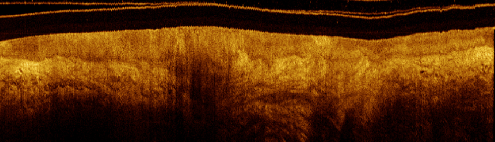


Optical coherence tomography (OCT) is the optical analog of ultrasonography (“ultrasound”). It enables the high-resolution examination of sub-surface tissue morphology and function in real-time. A one-dimensional OCT image (axial line or “A-line”) can be conceptually thought of as illuminating tissue with pulses of light and measuring the time required to detected reflected pulses from sub-surface surfaces within the tissue. The time-of-flight for each detected pulse is used to construct the A-line.
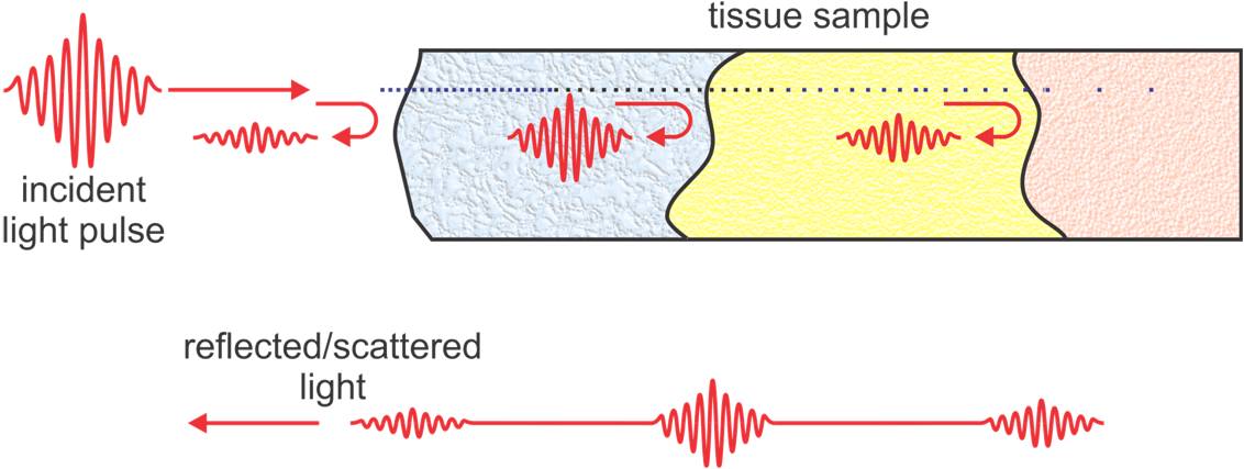
Owing to the very high speed of light – light travels more than 100,000 times faster than sound in tissue – time-of-flight cannot be measured directly. In OCT, an interferometer is employed to measure time-of-flight in either the time or frequency-domain. Two- and three-dimensional images can be constructed by scanning the imaging beam over the tissue surface.
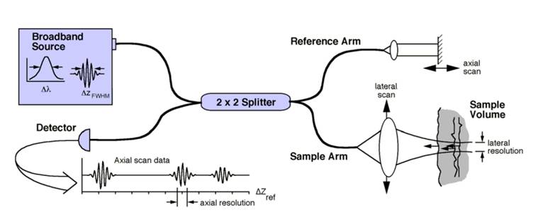
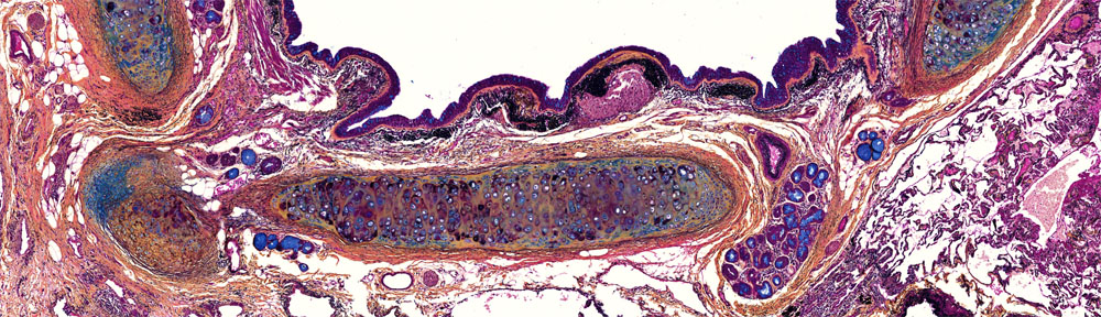
The resolution of OCT in the A-line direction is determined by the center wavelength and bandwidth of the laser source. Using a 1310 nm source with 100nm bandwidth, the A-line resolution is about 10 µm, sufficient for resolving tissue layers and larger morphological structures. Imaging depth in OCT is limited by the optical transparency (absorption and scattering) of the tissue but is typically on the order of 1 to 2 mm.
Rotary-Pullback Catheters
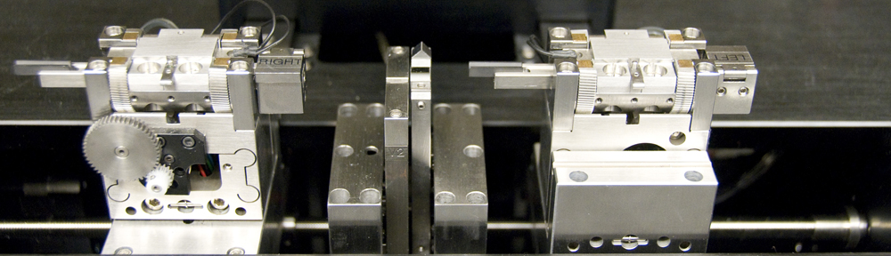
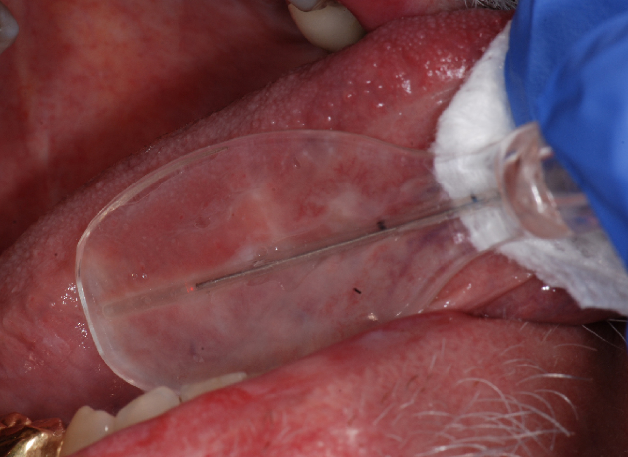
Accessing internal organs requires fiber-optic imaging catheters that are small enough to be inserted into the instrument channel of an endoscope for guidance to the desired imaging site. We have developed a rotary-pullback catheter for in vivo OCT imaging. A stainless steel torque cable drives the rotating fiber-optic core to sweep out circular scans at the distal tip of the catheter. The catheter is slightly forward-looking to enable Doppler sensitivity. The optical core is pulled back during rotation – sweeping out a spiral scan– to enable volumetric imaging.
In our catheter design, light is guided through a fiber-optic assembly consisting of a length of single-mode fiber (SMF) spliced to beam-shaping optics. The beam-shaping optics consists of a segment of no-core fiber (NCF), a segment of graded-index (GRIN) fiber to provide optical power (the lens), and a segment of angle-polished NCF to redirect the beam.
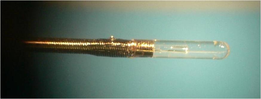
The first segment of NCF increases the fiber-to-GRIN distance to reduce the size of the projected spot (while increasing its NA) to improve lateral resolution while maintaining a long working distance. The GRIN segment focuses the OCT beam into the tissue while the angle-polished NCF employs total internal reflection to redirect the beam to the side of the fiber-optic assembly in order to capture cross-sectional images.
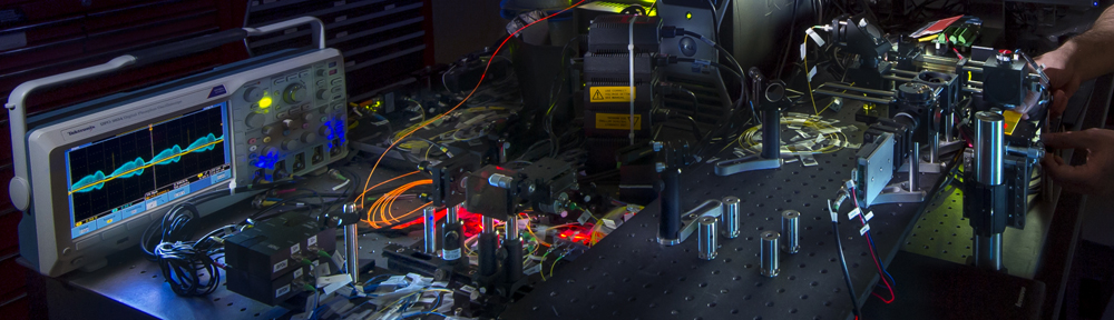
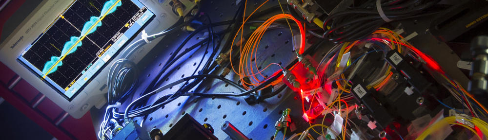
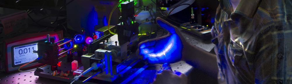
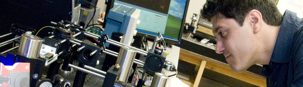
OCT Instrumentation
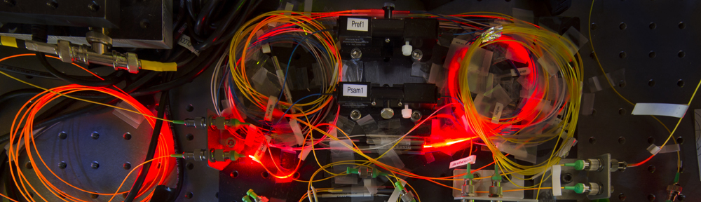
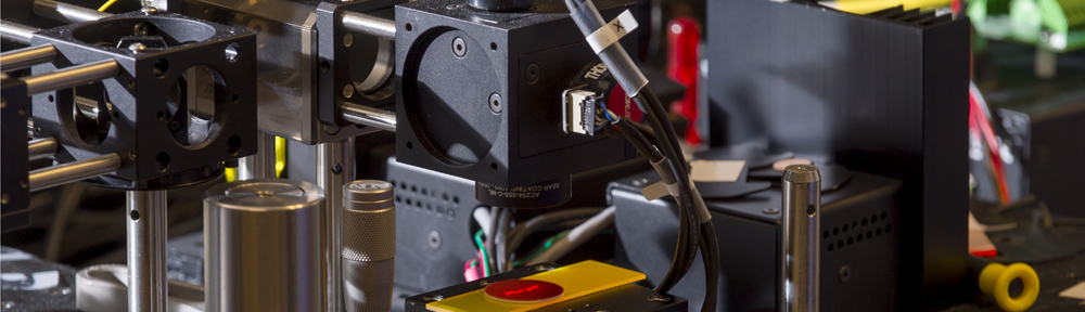
We develop our own Fourier-domain systems based on swept-source lasers for versatility and fast imaging. Custom instrumentation, rotary-pullback catheters, rotary-pullback drives, and data acquisition software allow us to extend the capabilities of OCT beyond purely structural imaging. A schematic of our OCT instrument is shown in the figure below. Our systems operate at 1310 nm striking a balance between shorter wavelengths where resolution is higher and longer wavelengths where tissue penetration better due to lower absorption by tissue constituents. Conveniently, this wavelength allows the use of readily available telecom fiber components to construct relatively inexpensive fiber optic OCT interferometers to detect the light returning from the tissue.
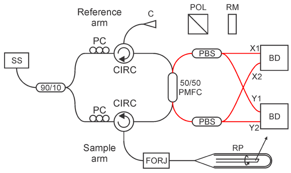
The rotary-pullback drive (RPD) actuates catheter rotation and pullback from the proximal end of the catheter. Rotational speeds up to 6000 rpm are possible enabling a maximum B-scan rate of 100Hz. Pullback speeds up to 15 mm/s are also selectable. Faster rotary and pullback speeds can be used for rapid volumetric imaging to limit motion artifacts while slower speeds can be used to create higher resolution images or for enabling OCT imaging extensions such as Doppler.
Continue with Autofluorescence Imaging (AFI).