Journal Articles
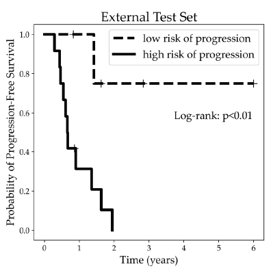
Janzen, Ian; Ho, Cheryl; Melosky, Barbara; Ye, Qian; Li, Jessica; Wang, Gang; Lam, Stephen; MacAulay, Calum; Yuan, Ren
In: MDPI Cancers, vol. 17, iss. 1, no. 58, 2024.
@article{Janzen2024,

title = {Machine Learning and Computed Tomography Radiomics to Predict Disease Progression to Upfront Pembrolizumab Monotherapy in Advanced Non-Small-Cell Lung Cancer: A Pilot Study},

author = {Ian Janzen AND Cheryl Ho AND Barbara Melosky AND Qian Ye AND Jessica Li AND Gang Wang AND Stephen Lam AND Calum MacAulay AND Ren Yuan},

url = {https://doi.org/10.3390/cancers17010058, DOI

https://biophotonics.bccrc.ca/wp-content/uploads/2025/01/cancers-17-00058-v2.pdf, PDF},

year  = {2024},

date = {2024-12-28},

urldate = {2024-12-28},

journal = {MDPI Cancers},

volume = {17},

number = {58},

issue = {1},

abstract = {Background/Objectives: Pembrolizumab monotherapy is approved in Canada for first-line treatment of advanced NSCLC with PD-L1 ≥ 50% and no EGFR/ALK aberrations. However, approximately 55% of these patients do not respond to pembrolizumab, underscoring the need for the early intervention of non-responders to optimize treatment strategies. Distinguishing the 55% sub-cohort prior to treatment is a real-world dilemma. Methods: In this retrospective study, we analyzed two patient cohorts treated with pembrolizumab monotherapy (training set: n = 97; test set: n = 17). The treatment response was assessed using baseline and follow-up CT scans via RECIST 1.1 criteria. Results: A logistic regression model, incorporating pre-treatment CT radiomic features of lung tumors and clinical variables, achieved high predictive accuracy (AUC: 0.85 in training; 0.81 in testing, 95% CI: 0.63–0.99). Notably, radiomic features from the peritumoral region were found to be independent predictors, complementing the standard CT evaluations and other clinical characteristics. Conclusions: This pragmatic model offers a valuable tool to guide first-line treatment decisions in NSCLC patients with high PD-L1 expression and has the potential to advance personalized oncology and improve timely disease management.},

keywords = {},

pubstate = {published},

tppubtype = {article}

}

Malone, Jeanie; Tanskanen, Adrian; Hill, Chloe; Cynamon, Allan Zuckermann; Hoang, Lien; MacAulay, Calum; McAlpine, Jessica N.; Lane, Pierre M.
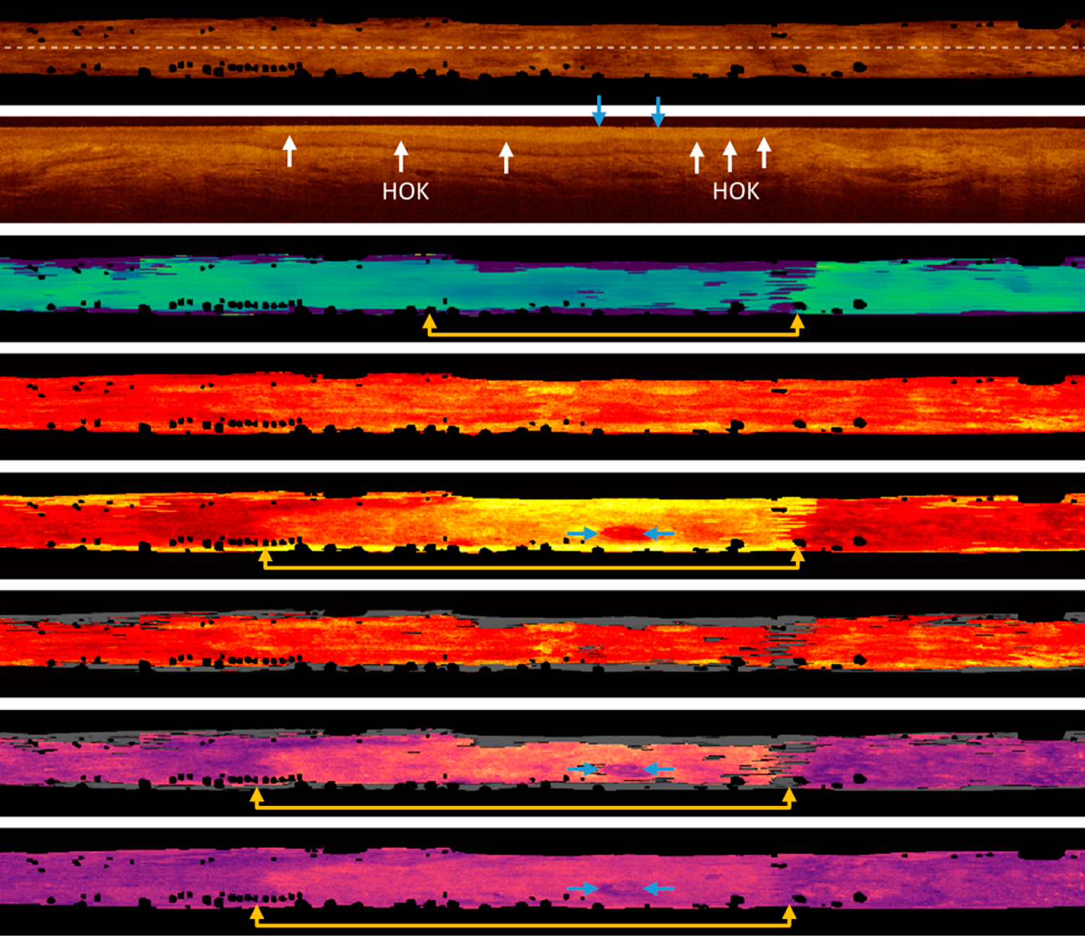
Multimodal Optical Imaging of Ex Vivo Fallopian Tubes to Distinguish Early and Occult Tubo-Ovarian Cancers Journal Article
In: MDPI Cancers, vol. 16, iss. 21, no. 3618, 2024.
@article{MaloneFallopian2024,

title = {Multimodal Optical Imaging of Ex Vivo Fallopian Tubes to Distinguish Early and Occult Tubo-Ovarian Cancers},

author = {Jeanie Malone AND Adrian Tanskanen AND Chloe Hill AND Allan Zuckermann Cynamon AND Lien Hoang AND Calum MacAulay AND Jessica N. McAlpine AND Pierre M. Lane

},

url = {https://doi.org/10.3390/cancers16213618, DOI,

https://biophotonics.bccrc.ca/wp-content/uploads/2024/10/cancers-16-03618.pdf, PDF},

year  = {2024},

date = {2024-10-26},

urldate = {2024-10-26},

journal = {MDPI Cancers},

volume = {16},

number = {3618},

issue = {21},

abstract = {Tubo-ovarian cancers are associated with high mortality as they are often not detected until a late stage. Early diagnosis is associated with better patient outcomes, but there are currently no effective screening measures. We explore whether an optical imaging catheter can detect early or occult lesions in the fallopian tubes. This device collects three-dimensional structural images of tissue through optical coherence tomography (OCT) simultaneously with functional imaging through autofluorescence imaging (AFI). We image ex vivo fallopian tubes from n = 28 patients (n = 7 cancer patients) and explore eleven imaging biomarkers for their ability to distinguish early or occult disease. We find that high-grade serous ovarian carcinomas can be visually distinguished through this approach, and that there are several quantitative changes within the area of lesion and throughout the specimen that can be measured through these imaging biomarkers. We conclude that this approach shows promise and merits further investigation of its diagnostic potential.},

keywords = {},

pubstate = {published},

tppubtype = {article}

}
Malone, Jeanie; Hill, Chloe; Tanskanen, Adrian; Liu, Kelly; Ng, Samson; MacAulay, Calum; Poh, Catherine F.; Lane, Pierre M.
Imaging Biomarkers of Oral Dysplasia and Carcinoma Measured with In Vivo Endoscopic Optical Coherence Tomography Journal Article
In: MDPI Cancers, vol. 16, iss. 15, no. 15, 2024.
@article{MaloneOral2024,

title = {Imaging Biomarkers of Oral Dysplasia and Carcinoma Measured with In Vivo Endoscopic Optical Coherence Tomography},

author = {Jeanie Malone AND Chloe Hill AND Adrian Tanskanen AND Kelly Liu AND Samson Ng AND Calum MacAulay AND Catherine F. Poh AND Pierre M. Lane},

url = {https://doi.org/10.3390/cancers16152751, DOI

https://biophotonics.bccrc.ca/wp-content/uploads/2024/08/cancers-16-02751.pdf, PDF},

year  = {2024},

date = {2024-08-02},

urldate = {2024-08-02},

journal = {MDPI Cancers},

volume = {16},

number = {15},

issue = {15},

abstract = {Oral cancers are associated with high mortality in advanced stages. Early diagnosis is associated with better patient outcomes, but this is challenging to achieve as benign lesions look similar to lesions of concern, and multiple biopsies may be required to ensure the most pathologic tissue is sampled. This work leverages a previously developed endoscopic imaging system and deep learning segmentation tool to provide measurements of subsurface changes in the first few millimeters of oral tissue. We present seven quantitative features that allow for rapid examination of tissue, which we propose may be useful for biopsy site or treatment margin selection.},

keywords = {},

pubstate = {published},

tppubtype = {article}

}
Hill, Chloe; Malone, Jeanie; Liu, Kelly; Ng, Samson Pak-Yan; MacAulay, Calum; Poh, Catherine; Lane, Pierre
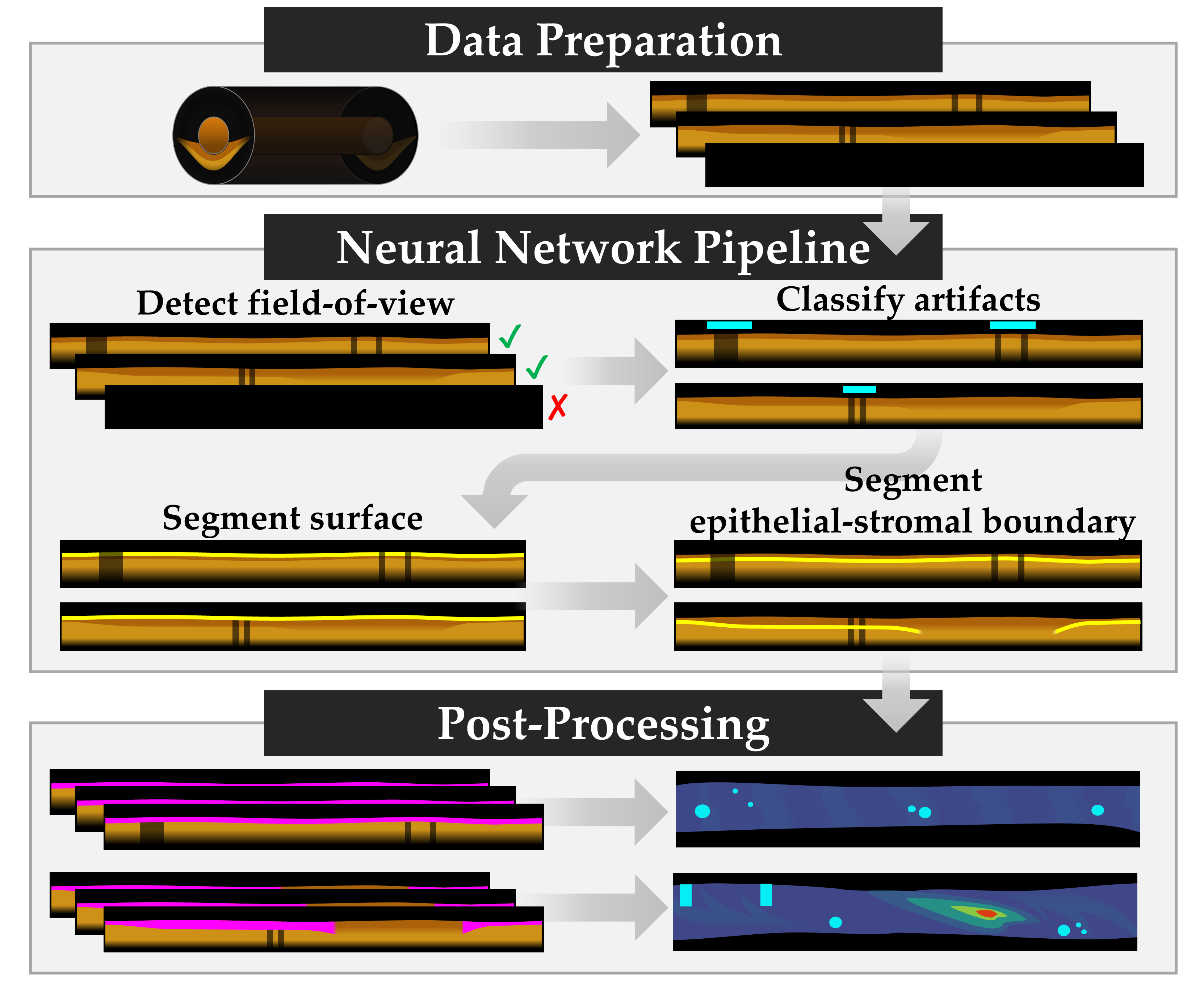
Three-Dimension Epithelial Segmentation in Optical Coherence Tomography of the Oral Cavity Using Deep Learning Journal Article
In: MDPI Cancers, vol. 16, iss. 11, pp. 2144, 2024.
@article{Hill2024,

title = {Three-Dimension Epithelial Segmentation in Optical Coherence Tomography of the Oral Cavity Using Deep Learning},

author = {Chloe Hill and Jeanie Malone and Kelly Liu and Samson Pak-Yan Ng and Calum MacAulay and Catherine Poh and Pierre Lane},

url = {https://doi.org/10.3390/cancers16112144, DOI

https://biophotonics.bccrc.ca/wp-content/uploads/2024/06/Hill-2024.pdf, PDF},

year  = {2024},

date = {2024-06-05},

urldate = {2024-06-05},

journal = {MDPI Cancers},

volume = {16},

issue = {11},

pages = {2144},

abstract = {This paper aims to simplify the application of optical coherence tomography (OCT) for the examination of subsurface morphology in the oral cavity and reduce barriers towards the adoption of OCT as a biopsy guidance device. The aim of this work was to develop automated software tools for the simplified analysis of the large volume of data collected during OCT. Imaging and corresponding histopathology were acquired in-clinic using a wide-field endoscopic OCT system. An annotated dataset (n = 294 images) from 60 patients (34 male and 26 female) was assembled to train four unique neural networks. A deep learning pipeline was built using convolutional and modified u-net models to detect the imaging field of view (network 1), detect artifacts (network 2), identify the tissue surface (network 3), and identify the presence and location of the epithelial–stromal boundary (network 4). The area under the curve of the image and artifact detection networks was 1.00 and 0.94, respectively. The Dice similarity score for the surface and epithelial–stromal boundary segmentation networks was 0.98 and 0.83, respectively. Deep learning (DL) techniques can identify the location and variations in the epithelial surface and epithelial–stromal boundary in OCT images of the oral mucosa. Segmentation results can be synthesized into accessible en face maps to allow easier visualization of changes.},

keywords = {},

pubstate = {published},

tppubtype = {article}

}
Searles, Kyle; Shalabi, Nabil; Hohert, Geoffrey; Gharib, Nirvana; Jayhooni, Sayed Mohammad Hashem; andKenichi Takahata, Pierre M. Lane
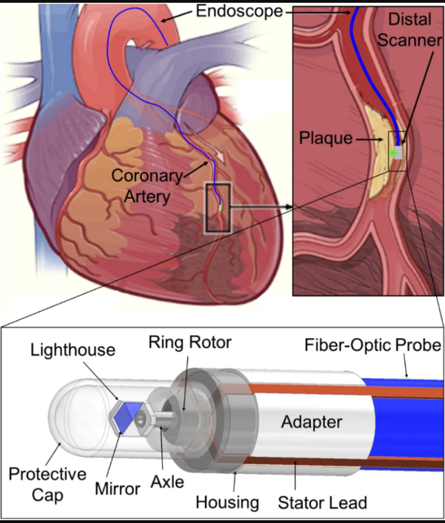
Distal planar rotary scanner for endoscopic optical coherence tomography Journal Article
In: Biomedical Engineering Letters, 2024.
@article{Searles2024,

title = {Distal planar rotary scanner for endoscopic optical coherence tomography},

author = {Kyle Searles and Nabil Shalabi and Geoffrey Hohert and Nirvana Gharib and Sayed Mohammad Hashem Jayhooni and Pierre M. Lane andKenichi Takahata},

url = {https://doi.org/10.1007/s13534-024-00353-8, DOI

https://biophotonics.bccrc.ca/wp-content/uploads/2024/02/s13534-024-00353-8.pdf, Full Text (PDF)},

year  = {2024},

date = {2024-02-19},

urldate = {2024-02-19},

journal = {Biomedical Engineering Letters},

abstract = {Optical coherence tomography (OCT) is becoming a more common endoscopic imaging modality for detecting and treating disease given its high resolution and image quality. To use OCT for 3-dimensional imaging of small lumen, embedding an optical scanner at the distal end of an endoscopic probe for circumferential scanning the probing light is a promising way to implement high-quality imaging unachievable with the conventional method of revolving an entire probe. To this end, the present work proposes a hollow and planar micro rotary actuator for its use as an endoscopic distal scanner. A miniaturized design of this ferrofluid-assisted electromagnetic actuator is prototyped to act as a full 360° optical scanner, which is integrated at the tip of a fiber-optic probe together with a gradient-index lens for use with OCT. The scanner is revealed to achieve a notably improved dynamic performance that shows a maximum speed of 6500 rpm, representing 325% of the same reported with the preceding design, while staying below the thermal limit for safe in-vivo use. The scanner is demonstrated to perform real-time OCT using human fingers as live tissue samples for the imaging tests. The acquired images display no shadows from the electrical wires to the scanner, given its hollow architecture that allows the probing light to pass through the actuator body, as well as the quality high enough to differentiate the dermis from the epidermis while resolving individual sweat glands, proving the effectiveness of the prototyped scanner design for endoscopic OCT application.},

keywords = {},

pubstate = {published},

tppubtype = {article}

}
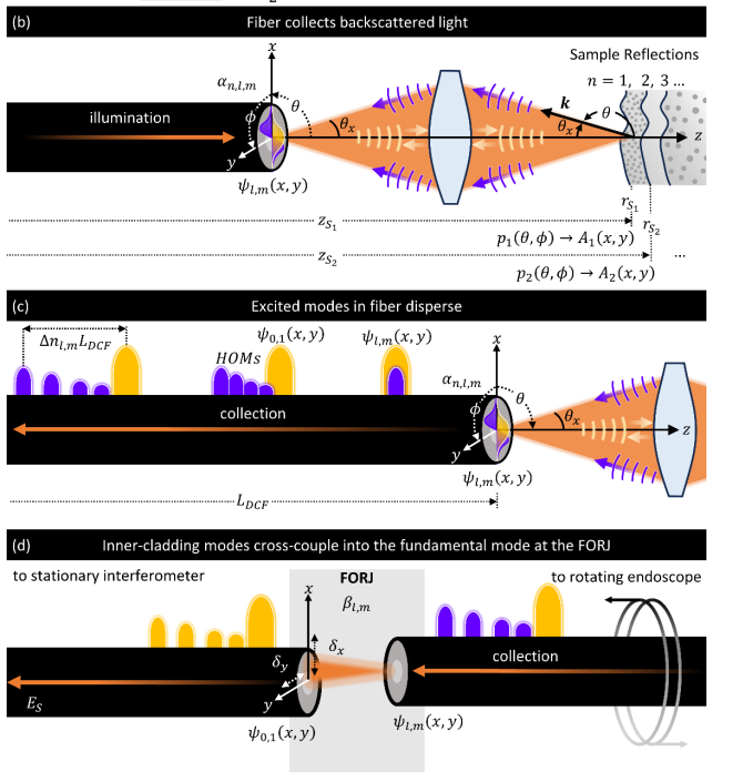
Tanskanen, Adrian; Malone, Jeanie; MacAulay, Calum; Lane, Pierre
Multipath artifacts enable angular contrast in multimodal endoscopic optical coherence tomography Journal Article
In: Optics Express Vol. 31, Issue 26, vol. 31, iss. 26, pp. 44224-44245, 2023.
@article{TanskanenMalone2023,

title = {Multipath artifacts enable angular contrast in multimodal endoscopic optical coherence tomography},

author = {Adrian Tanskanen and Jeanie Malone and Calum MacAulay and Pierre Lane},

url = {https://doi.org/10.1364/OE.504854, DOI

https://biophotonics.bccrc.ca/wp-content/uploads/2023/12/oe-31-26-44224.pdf, Full Text (PDF)},

year  = {2023},

date = {2023-12-12},

urldate = {2023-12-12},

journal = {Optics Express Vol. 31, Issue 26},

volume = {31},

issue = {26},

pages = {44224-44245},

abstract = {Multipath artifacts are inherent to double-clad fiber based optical coherence tomography (OCT), appearing as ghost images blurred in the A-line direction. They result from the excitation of higher-order inner-cladding modes in the OCT sample arm which cross-couple into the fundamental mode at discontinuities and thus are detected in single-mode fiber-based interferometers. Historically, multipath artifacts have been regarded as a drawback in single fiber endoscopic multimodal OCT systems as they degrade OCT quality. In this work, we reveal that multipath artifacts can be projected into high-quality two-dimensional en face images which encode high angle backscattering features. Using a combination of experiment and simulation, we characterize the coupling of Mie-range scatterers into the fundamental image (LP01 mode) and higher-order image (multipath artifact). This is validated experimentally through imaging of microspheres with an endoscopic multimodal OCT system. The angular dependence of the fundamental image and higher order image generated by the multipath artifact lays the basis for multipath contrast, a ratiometric measurement of differential coupling which provides information regarding the angular diversity of a sample. Multipath contrast images can be generated from OCT data where multipath artifacts are present, meaning that a wealth of clinical data can be retrospectively examined.},

keywords = {},

pubstate = {published},

tppubtype = {article}

}
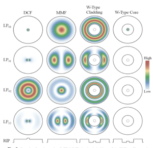
Tanskanen, Adrian; Malone, Jeanie; Hohert, Geoffrey; MacAulay, Calum; Lane, Pierre
Triple-clad W-type fiber mitigates multipath artifacts in multimodal optical coherence tomography Journal Article
In: Optics Express, vol. 31, pp. 4465-4481, 2023.
@article{Tanskanen2023,

title = {Triple-clad W-type fiber mitigates multipath artifacts in multimodal optical coherence tomography},

author = {Adrian Tanskanen and Jeanie Malone and Geoffrey Hohert and Calum MacAulay and Pierre Lane},

url = {https://doi.org/10.1364/OE.476768, DOI

https://biophotonics.bccrc.ca/wp-content/uploads/2023/01/oe-31-3-4465.pdf, Full Text (PDF)

},

year  = {2023},

date = {2023-01-24},

urldate = {2023-01-24},

journal = {Optics Express},

volume = {31},

pages = {4465-4481},

abstract = {Multimodal endoscopic optical coherence tomography (OCT) can be implemented with double-clad fiber by using the presumed single-mode core for OCT and the higher numerical aperture cladding for a secondary modality. However, the quality of OCT in double-clad fiber (DCF) based systems is compromised by the introduction of multipath artifacts that aren't present in single-mode fiber OCT systems. Herein, the mechanisms for multipath artifacts in DCF are linked to its modal contents using a commercial software package and experimental measurement. A triple-clad W-type fiber is proposed as a method for achieving multimodal imaging with single-mode quality OCT in an endoscopic system. Simulations of the modal contents of a W-type fiber are compared to DCF and single-mode fiber. Finally, a W-Type fiber rotary catheter is used in a DCF-based endoscopic OCT and autofluorescence imaging (AFI) system to demonstrate multipath artifact free OCT and AFI of a human fingertip.},

keywords = {},

pubstate = {published},

tppubtype = {article}

}
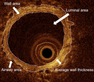
Peters, Carli M.; Peters, Robert C.; Lee, Anthony D.; Lane, Pierre; Lam, Stephen; Sin, Don D.; McKenzie, Donald C.; Sheel, A. William
Software development to optimize the minimal detectable difference in human airway images captured using optical coherence tomography Journal Article
In: Clinical Physiology and Functional Imaging, iss. 42, pp. 308–319, 2022.
@article{Peters2022,

title = {Software development to optimize the minimal detectable difference in human airway images captured using optical coherence tomography},

author = {Carli M. Peters and Robert C. Peters and Anthony D. Lee and Pierre Lane and Stephen Lam and Don D. Sin and Donald C. McKenzie and A. William Sheel},

url = {https://doi.org/10.1111/cpf.12762, DOI

https://biophotonics.bccrc.ca/wp-content/uploads/2023/01/Clin-Physio-Funct-Imaging-2022-Peters-Software-development-to-optimize-the-minimal-detectable-difference-in-human.pdf, Full Text (PDF)},

year  = {2022},

date = {2022-05-04},

urldate = {2022-05-04},

journal = {Clinical Physiology and Functional Imaging},

issue = {42},

pages = {308–319},

abstract = {Optical coherence tomography (OCT) is an imaging methodology that can be used to assess human airways. OCT avoids the harmful effects of ionizing radiation and has a high spatial resolution making it well suited for imaging the structure of small airways. Analysis of OCT airway images has typically been performed manually by tracing the airway with a relatively high coefficient of variation. The purpose of this study was to develop an analysis tool to reduce the inter‐ and intra‐observer reproducibility of OCT and improve the ability to detect differences in airways. OCT images from healthy, young human volunteers were used to develop and test the OCT software. Measurement software was developed to allow the conversion of the original image into a grayscale image and was followed by an enhancement operation to brighten the image, and contour measurement. A total of 140 OCT images, 70 small (<2 mm) and 70 medium (2–4 mm) sized airways were analyzed. The inter‐ and intraobserver reproducibility of airway measurements ranged for strong to very strong in the small‐sized airways. For medium‐sized airways the reproducibility was considered moderate. Bland‐Altman bias was low between observers and observations for all measures. The minimal detectable differences in the airway measurements with our semi‐automated software were lower relative to manual tracing in medium‐sized airways. Our software improves the ability to perform quantitative OCT analysis and may help to quantify the extent of airway remodelling in respiratory disease or elite athletes in future studies.},

keywords = {},

pubstate = {published},

tppubtype = {article}

}
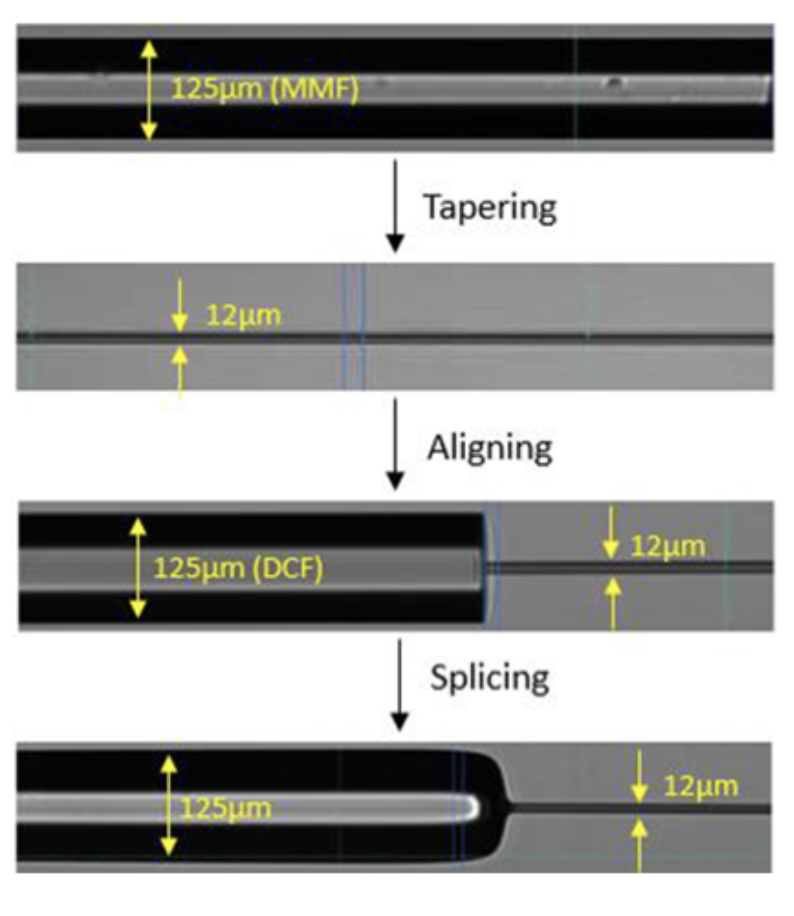
Kaur, Mandeep; Hohert, Geoffrey; Lane, Pierre M.; Menon, Carlo
Fabrication of a stepped optical fiber tip for miniaturized scanners Journal Article
In: Optical Fiber Technology, vol. 61, no. 102436, 2021.
@article{Kaur2021,

title = {Fabrication of a stepped optical fiber tip for miniaturized scanners},

author = {Mandeep Kaur and Geoffrey Hohert and Pierre M. Lane and Carlo Menon},

url = {https://doi.org/10.1016/j.yofte.2020.102436, DOI

https://biophotonics.bccrc.ca/wp-content/uploads/2022/02/1-s2.0-S1068520020304260-main.pdf, Full Text (PDF)},

year  = {2021},

date = {2021-12-30},

urldate = {2021-12-30},

journal = {Optical Fiber Technology},

volume = {61},

number = {102436},

abstract = {Advancements in fabrication of miniaturized optical scanners would benefit from micrometer sized optical fiber tips. The change in the cross section of an optical fiber tip is often accompanied with the presence of a longer tapered area. The reduction of the cross section of double clad optical fibers (DCFs) with a flat interface surface at the region where a change in the cross section takes place (with an abrupt change in the cross section) is considered in this paper. Various methods such as heating and pulling, wet etching using hydrofluoric acid (HF), and etching in a vaporous state were explored. The optical etching rate and its dependence on the temperature of the etchant solution were also determined. Optical fibers etch linearly with time, and the etching speed is dependent on the temperature of the etchant solution which shows a parabolic trend. The flatness of the surface at the cross section change is an interesting parameter in the fabrication of submillimeter sized scanners where the light is transmitted through the core of the DCF, and reflected light is collected through the inner cladding of the same fiber, or vice versa. The surface flatness at the interface was compared among different fiber samples developed using the aforementioned techniques. This research illustrates that the wet chemical etching performed by blocking the capillary rising of etchant solution along the fiber provided advantages over the heating and pulling technique in terms of light intensity transmitted to the target sample and the reflected light collected through the interface of etched cladding.},

keywords = {},

pubstate = {published},

tppubtype = {article}

}
Peters, Carli M.; Leahy, Michael G.; Hohert, Geoffrey; Lane, Pierre; Lam, Stephen; Sin, Don D.; McKenzie, Donald C.; Sheel, Andrew William
Airway luminal area and the resistive work of breathing during exercise in healthy young females and males Journal Article
In: J Appl Physiol, vol. 131, iss. 6, pp. 1750-1761, 2021.
@article{Peters2021,

title = {Airway luminal area and the resistive work of breathing during exercise in healthy young females and males},

author = {Carli M. Peters and Michael G. Leahy and Geoffrey Hohert and Pierre Lane and Stephen Lam and Don D. Sin and Donald C. McKenzie and Andrew William Sheel },

url = {https://doi.org/10.1152/japplphysiol.00418.2021, DOI},

year  = {2021},

date = {2021-12-01},

urldate = {2021-12-01},

journal = {J Appl Physiol},

volume = {131},

issue = {6},

pages = {1750-1761},

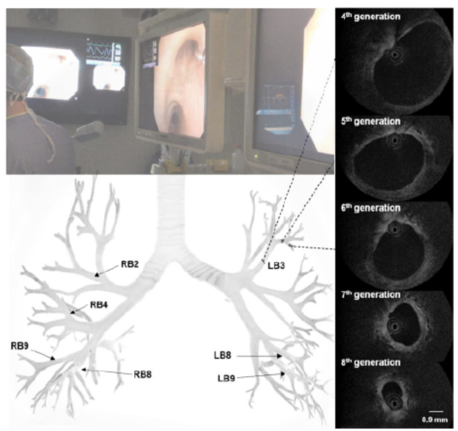
abstract = {We examined the relationship between the work of breathing (Wb) during exercise and in vivo measures of airway size in healthy females and males. We hypothesized that sex differences in airway luminal area would explain the larger resistive Wb during exercise in females. Healthy participants (n = 11 females and n = 11 males; 19-30 yr) completed a cycle exercise test to exhaustion where Wb was assessed using an esophageal balloon catheter. On a separate day, each participant underwent a bronchoscopy procedure for optical coherence tomography measures of seven airways. In vivo measures of luminal area were made for the fourth to eighth airway generations. A composite index of airway size was calculated as the sum of the luminal area for each generation, and the total area was calculated based on Weibel's model. We found that index of airway size (males: 37.4 ± 6.3 mm2 vs. females: 27.5 ± 7.4 mm2) and airway area calculated based on Weibel's model (males: 2,274 ± 557 mm2 vs. females: 1,594 ± 389 mm2) were significantly larger in males (both P = 0.003). When minute ventilation was greater than ∼60 L·min-1, the resistive Wb was higher in females. At the highest equivalent flow achieved by all subjects, resistance to inspired flow was larger in females and significantly associated with two measures of airway size in all subjects: index of airway size (r = 0.524, P = 0.012) and Weibel area (r = 0.525, P = 0.012). Our findings suggest that innate sex differences in luminal area result in a greater resistive Wb during exercise in females compared with males.NEW & NOTEWORTHY We hypothesized that the higher resistive work of breathing in females compared with males during high-intensity exercise is due to smaller airways. In vivo measures of the fourth to eighth airway generations made using optical coherence tomography show that females tend to have smaller airway luminal areas of the fourth to sixth airway generations. Sex differences in airway luminal area result in a greater resistive work of breathing during exercise in females compared with males.},

keywords = {},

pubstate = {published},

tppubtype = {article}

}
Tanskanen, Adrian; Hohert, Geoffrey; Lee, Anthony; Lane, Pierre M.
Higher-Order Core-Like Modes in Double-Clad Fiber Contribute to Multipath Artifacts in Optical Coherence Tomography Journal Article
In: Journal of Lightwave Technology, vol. 39, no. 17, pp. 5573-5581, 2021.
@article{Tanskanen2021,

title = {Higher-Order Core-Like Modes in Double-Clad Fiber Contribute to Multipath Artifacts in Optical Coherence Tomography},

author = {Adrian Tanskanen and Geoffrey Hohert and Anthony Lee and Pierre M. Lane},

url = {https://doi.org/10.1109/JLT.2021.3088055, DOI},

year  = {2021},

date = {2021-09-01},

urldate = {2021-09-01},

journal = {Journal of Lightwave Technology},

volume = {39},

number = {17},

pages = {5573-5581},

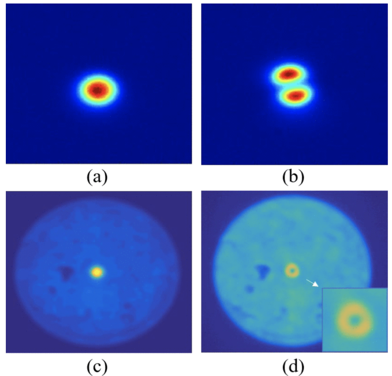
abstract = {Double-clad fiber (DCF) has enabled the combination of endoscopic optical coherence tomography (OCT) with secondary optical modalities. While DCF offers an additional optical channel, it is widely understood that its use reduces the quality of OCT owing to the introduction of multipath artifacts. We show here that an unexpected higher-order mode (HOM) with its energy confined to the DCF core can contribute to these artifacts. The existence of this HOM is confirmed using the spatially and spectrally (S 2 ) resolved imaging method. The group delay difference of the HOM is shown to be consistent with the delay of the diffuse ghost artifact in DCF-acquired OCT images.},

keywords = {},

pubstate = {published},

tppubtype = {article}

}
Malone, Jeanie; Lee, Anthony M. D.; Hohert, Geoffrey; Nador, Roland G.; Lane, Pierre
Small airway dilation measured by endoscopic optical coherence tomography correlates with chronic lung allograft dysfunction Journal Article
In: Journal of Biomedical Optics , vol. 26, iss. 7, no. 076005, 2021.
@article{Malone2021,

title = {Small airway dilation measured by endoscopic optical coherence tomography correlates with chronic lung allograft dysfunction},

author = {Jeanie Malone and Anthony M. D. Lee and Geoffrey Hohert and Roland G. Nador and Pierre Lane

},

url = {https://doi.org/10.1117/1.JBO.26.7.076005, DOI

https://biophotonics.bccrc.ca/wp-content/uploads/2022/02/076005_1.pdf, Full Text (PDF)},

year  = {2021},

date = {2021-07-14},

journal = {Journal of Biomedical Optics },

volume = {26},

number = {076005},

issue = {7},

abstract = {Significance: Chronic lung allograft dysfunction (CLAD) is the leading cause of death in transplant patients who survive past the first year post-transplant. Current diagnosis is based on sustained decline in lung function; there is a need for tools that can identify CLAD onset.


Aim: Endoscopic optical coherence tomography (OCT) can visualize structural changes in the small airways, which are of interest in CLAD progression. We aim to identify OCT features in the small airways of lung allografts that correlate with CLAD status.


Approach: Imaging was conducted with an endoscopic rotary pullback OCT catheter during routine bronchoscopy procedures (n  =  54), collecting volumetric scans of three segmental airways per patient. Six features of interest were identified, and four blinded raters scored the dataset on the presence and intensity of each feature.


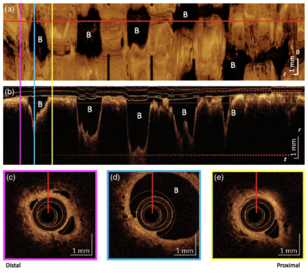
Results: Airway dilation (AD) was the only feature found to significantly (p  <  0.003) correlate with CLAD diagnosis (R  =  0.40 to 0.61). AD could also be fairly consistently scored between raters (κinter-rater  =  0.48, κintra-rater  =  0.64). There is a stronger relationship between AD and the combined obstructive and restrictive (BOS + RAS) phenotypes than the obstructive-only (BOS) phenotype for two raters (R  =  0.92  ,  0.94).


Conclusions: OCT examination of small AD shows potential as a diagnostic indicator for CLAD and CLAD phenotype and merits further exploration.},

keywords = {},

pubstate = {published},

tppubtype = {article}

}
Aim: Endoscopic optical coherence tomography (OCT) can visualize structural changes in the small airways, which are of interest in CLAD progression. We aim to identify OCT features in the small airways of lung allografts that correlate with CLAD status.
Approach: Imaging was conducted with an endoscopic rotary pullback OCT catheter during routine bronchoscopy procedures (n = 54), collecting volumetric scans of three segmental airways per patient. Six features of interest were identified, and four blinded raters scored the dataset on the presence and intensity of each feature.
Results: Airway dilation (AD) was the only feature found to significantly (p < 0.003) correlate with CLAD diagnosis (R = 0.40 to 0.61). AD could also be fairly consistently scored between raters (κinter-rater = 0.48, κintra-rater = 0.64). There is a stronger relationship between AD and the combined obstructive and restrictive (BOS + RAS) phenotypes than the obstructive-only (BOS) phenotype for two raters (R = 0.92 , 0.94).
Conclusions: OCT examination of small AD shows potential as a diagnostic indicator for CLAD and CLAD phenotype and merits further exploration.
Jayhooni, Sayed Mohammad Hashem; Hohert, Geoffrey; Assadsangabi, Babak; Lane, Pierre M.; Zeng, Haishan; Takahata, Kenichi
A Side-Viewing Endoscopic Probe With Distal Micro Rotary Scanner for Multimodal Luminal Imaging and Analysis Journal Article
In: Journal of Microelectromechanical Systems, vol. 30, no. 3, pp. 433-441, 2021.
@article{Jayhooni2021,

title = {A Side-Viewing Endoscopic Probe With Distal Micro Rotary Scanner for Multimodal Luminal Imaging and Analysis},

author = {Sayed Mohammad Hashem Jayhooni and Geoffrey Hohert and Babak Assadsangabi and Pierre M. Lane and Haishan Zeng and Kenichi Takahata},

url = {https://doi.org/10.1109/JMEMS.2021.3072617, DOI},

year  = {2021},

date = {2021-06-01},

urldate = {2021-06-01},

journal = {Journal of Microelectromechanical Systems},

volume = {30},

number = {3},

pages = {433-441},

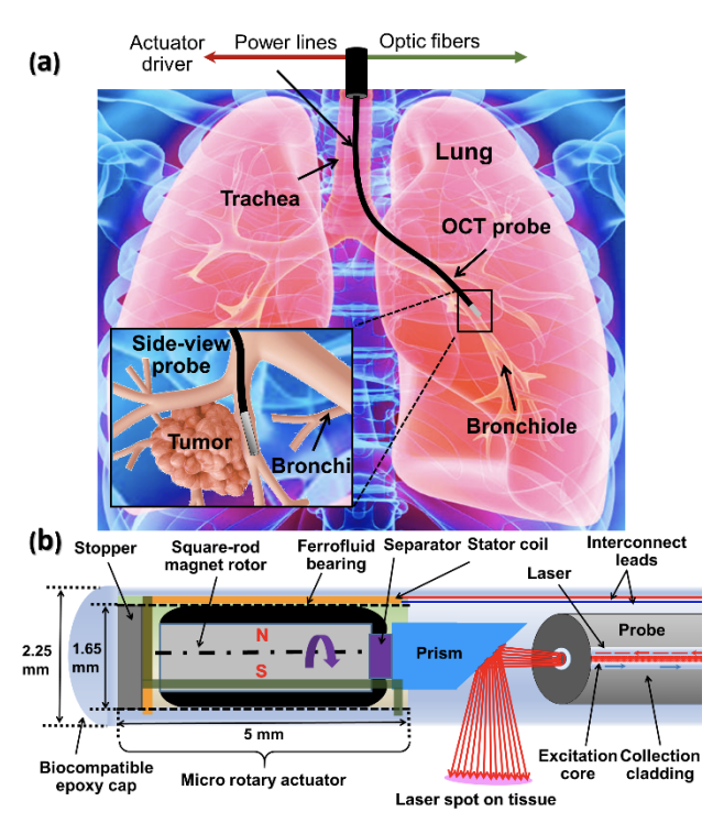
abstract = {This paper reports a novel micro electromagnetic actuator serving as the distal rotary scanner of an endoscopic probe developed for side-viewing luminal tissue screening and analysis enabled through different endoscopic modalities. The micro rotary optical scanner, for the first time, offers stepwise, low-speed, and high-speed rotational motions to be compatible with endoscopic optical coherence tomography (OCT) and Raman spectroscopy. In comparison with preceding designs, this rotary scanner is shown to provide up to 125× higher revolution speeds per power via a ~50% narrower body while limiting its operation temperature within a biologically safe level for in-vivo scanning purposes. A preliminary experiment on human skin tissues is performed using the prototyped side-viewing OCT endoscopic probe equipped with the developed distal scanner. The results demonstrate the probe's ability for real-time 360° imaging of live tissue with both high speed and high resolution. These results pave the path for further testing on human internal lumens.},

keywords = {},

pubstate = {published},

tppubtype = {article}

}
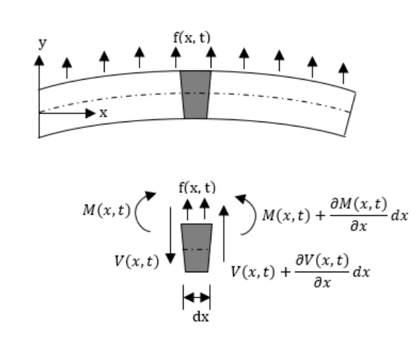
Kaur, Mandeep; Lane, Pierre M.; Menon, Carlo
Scanning and Actuation Techniques for Cantilever-Based Fiber Optic Endoscopic Scanners—A Review Journal Article
In: Sensors, vol. 21, iss. 1, no. 251, 2021.
@article{Kaur2021b,

title = {Scanning and Actuation Techniques for Cantilever-Based Fiber Optic Endoscopic Scanners—A Review},

author = {Mandeep Kaur and Pierre M. Lane and Carlo Menon},

url = {https://doi.org/10.3390/s21010251, DOI

https://biophotonics.bccrc.ca/wp-content/uploads/2022/02/sensors-21-00251-v2.pdf, Full Text (PDF)},

year  = {2021},

date = {2021-01-02},

urldate = {2021-01-02},

journal = {Sensors},

volume = {21},

number = {251},

issue = {1},

abstract = {Endoscopes are used routinely in modern medicine for in-vivo imaging of luminal organs. Technical advances in the micro-electro-mechanical system (MEMS) and optical fields have enabled the further miniaturization of endoscopes, resulting in the ability to image previously inaccessible small-caliber luminal organs, enabling the early detection of lesions and other abnormalities in these tissues. The development of scanning fiber endoscopes supports the fabrication of small cantilever-based imaging devices without compromising the image resolution. The size of an endoscope is highly dependent on the actuation and scanning method used to illuminate the target image area. Different actuation methods used in the design of small-sized cantilever-based endoscopes are reviewed in this paper along with their working principles, advantages and disadvantages, generated scanning patterns, and applications.},

keywords = {},

pubstate = {published},

tppubtype = {article}

}
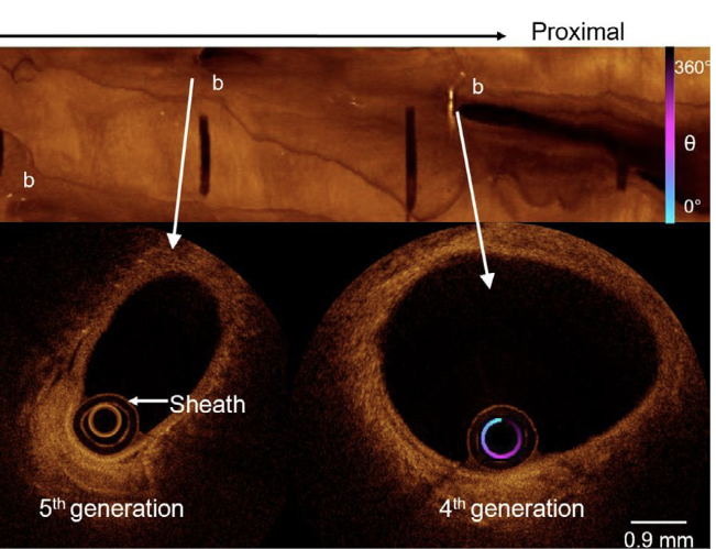
Peters, Carli M.; Molgat-Seon, Yannick; Dominelli, Paolo B.; Lee, Anthony M. D.; Lane, Pierre; Lam, Stephen; Sheel, Andrew W.
In: Physiological Reports, vol. 9, iss. 1, 2020.
@article{Peters2020,

title = {Fiber optic endoscopic optical coherence tomography (OCT) to assess human airways: The relationship between anatomy and physiological function during dynamic exercise},

author = {Carli M. Peters and Yannick Molgat-Seon and Paolo B. Dominelli and Anthony M. D. Lee and Pierre Lane and Stephen Lam and Andrew W. Sheel},

url = {https://biophotonics.bccrc.ca/wp-content/uploads/2022/02/Physiological-Reports-2021-Peters-Fiber-optic-endoscopic-optical-coherence-tomography-OCT-to-assess-human-airways-.pdf, Full Text (PDF)

https://doi.org/10.14814/phy2.14657, DOI},

year  = {2020},

date = {2020-12-28},

urldate = {2020-12-28},

journal = {Physiological Reports},

volume = {9},

issue = {1},

abstract = {Airway luminal area (Ai) influences respiratory mechanics during dynamic exercise; however, previous studies have investigated the relationship between airway anatomy and physiological function in different groups of individuals. The purpose of this study was to determine the effect of Ai on respiratory mechanics by making in vivo measures of airway dimensions and work of breathing (Wb) in the same individuals. Healthy participants (3F/2M; 23–45 years) completed a cycle exercise test to exhaustion. During exercise, Wb was assessed using an esophageal balloon catheter, while simultaneously assessing minute ventilation (urn:x-wiley:2051817X:media:phy214657:phy214657-math-0001E). On a separate day, subjects underwent a bronchoscopy procedure to capture optical coherence tomography (OCT) measures of three airways in the right lung. Each participant's Wb-urn:x-wiley:2051817X:media:phy214657:phy214657-math-0002E data were fit to a non-linear regression equation (Wb = aurn:x-wiley:2051817X:media:phy214657:phy214657-math-0003E3 + burn:x-wiley:2051817X:media:phy214657:phy214657-math-0004E2) that partitions Wb into its turbulent resistive (a) and viscoelastic (b) components. Measures of Ai and luminal diameter were made for the 4th–6th airway generations. A composite index of airway size was calculated as the sum of the Ai for each generation and the total area of the 4th–6th generation was calculated based on Weibel's model. Constant a was significantly correlated to the Weibel model total airway area (r = −0.94, p = 0.017) and index of airway size (r = −0.929, p = 0.023), whereas constant b was not associated with either measure (both p > 0.05). We found that individuals who had the smallest Ai had the highest resistive Wb and our findings provide the basis for further study of the relationship between airway size and respiratory mechanics during exercise.},

keywords = {},

pubstate = {published},

tppubtype = {article}

}
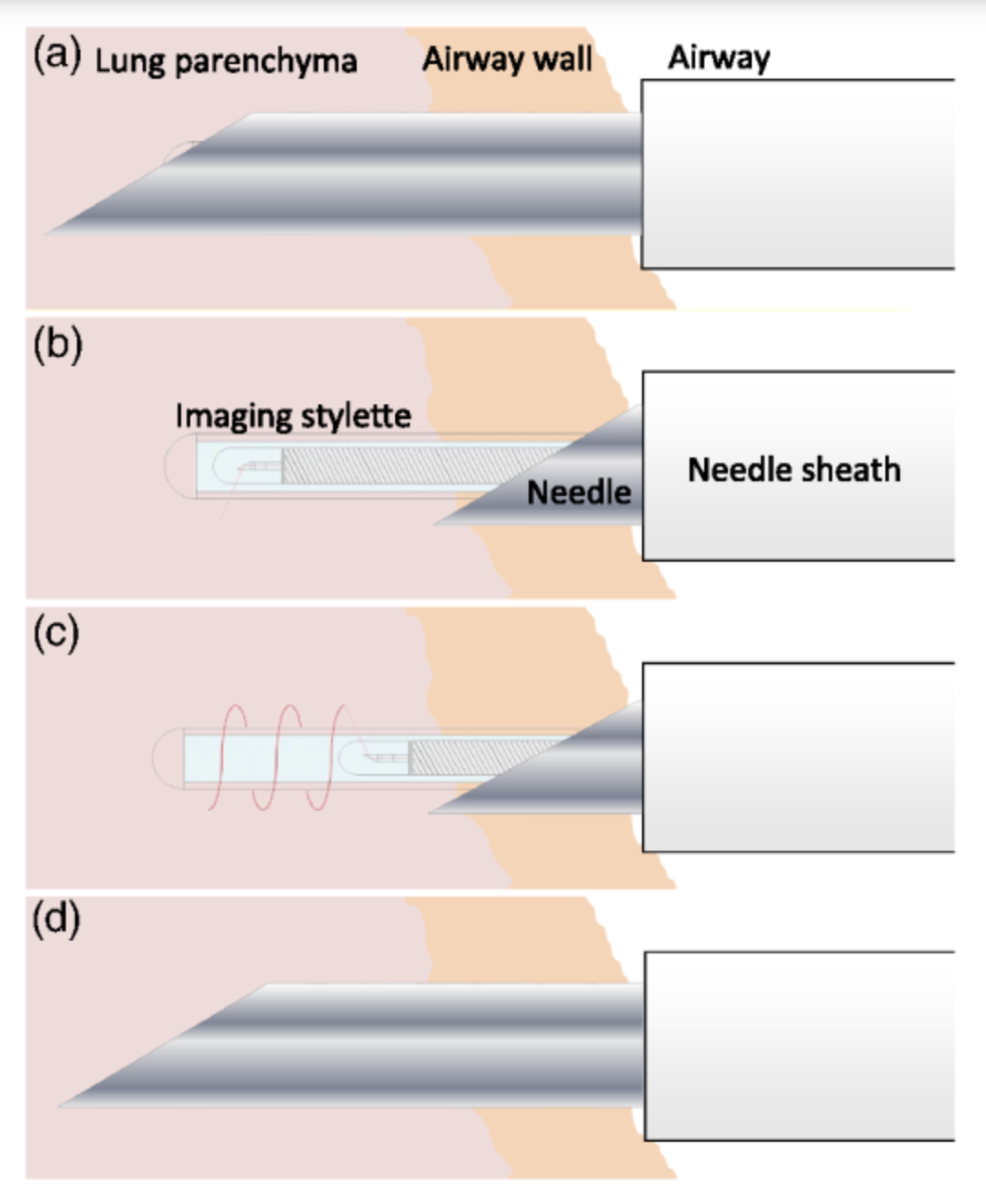
Hohert, Geoffrey; Myers, Renelle; Lam, Sylvia; Vertikov, Andrei; Lee, Anthony; Lam, Stephen; Lane, Pierre
Feasibility of combined optical coherence tomography and autofluorescence imaging for visualization of needle biopsy placement Journal Article
In: Journal of Biomedical Optics , vol. 25, iss. 10, no. 106003, 2020.
@article{Hohert2020,

title = {Feasibility of combined optical coherence tomography and autofluorescence imaging for visualization of needle biopsy placement},

author = {Geoffrey Hohert and Renelle Myers and Sylvia Lam and Andrei Vertikov and Anthony Lee and Stephen Lam and Pierre Lane},

url = {https://doi.org/10.1117/1.jbo.25.10.106003, DOI

https://biophotonics.bccrc.ca/wp-content/uploads/2022/02/106003_1.pdf, Full Text (PDF)},

year  = {2020},

date = {2020-10-20},

journal = {Journal of Biomedical Optics },

volume = {25},

number = {106003},

issue = {10},

abstract = {Significance: Diagnosis of suspicious lung nodules requires precise collection of relevant biopsies for histopathological analysis. Using optical coherence tomography and autofluorescence imaging (OCT-AFI) to improve diagnostic yield in parts of the lung inaccessible to larger imaging methods may allow for reducing complications related to the alternative of computed tomography-guided biopsy.


Aim: Feasibility of OCT-AFI combined with a commercially available lung biopsy needle was demonstrated for visualization of needle puncture sites in airways with diameters as small as 1.9 mm.


Approach: A miniaturized OCT-AFI imaging stylet was developed to be inserted through an 18G biopsy needle. We present design considerations and procedure development for image-guided biopsy. Ex vivo and in vivo porcine studies were performed to demonstrate the feasibility of the procedure and the device.


Results: OCT-AFI scans were obtained ex vivo and in vivo. Discrimination of pullback site is clear.


Conclusions: Use of the device is shown to be feasible in vivo. Images obtained show the stylet is effective at providing structural information at the puncture site that can be used to assess the diagnostic potential of the sample prior to collection.},

keywords = {},

pubstate = {published},

tppubtype = {article}

}
Aim: Feasibility of OCT-AFI combined with a commercially available lung biopsy needle was demonstrated for visualization of needle puncture sites in airways with diameters as small as 1.9 mm.
Approach: A miniaturized OCT-AFI imaging stylet was developed to be inserted through an 18G biopsy needle. We present design considerations and procedure development for image-guided biopsy. Ex vivo and in vivo porcine studies were performed to demonstrate the feasibility of the procedure and the device.
Results: OCT-AFI scans were obtained ex vivo and in vivo. Discrimination of pullback site is clear.
Conclusions: Use of the device is shown to be feasible in vivo. Images obtained show the stylet is effective at providing structural information at the puncture site that can be used to assess the diagnostic potential of the sample prior to collection.
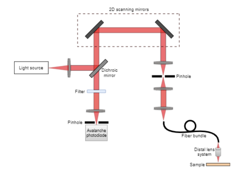
Kaur, Mandeep; Lane, Pierre M.; Menon, Carlo
Endoscopic Optical Imaging Technologies and Devices for Medical Purposes: State of the Art Journal Article
In: Applied Science, vol. 10, iss. 19, no. 6865, 2020.
@article{Kaur2020b,

title = {Endoscopic Optical Imaging Technologies and Devices for Medical Purposes: State of the Art},

author = {Mandeep Kaur and Pierre M. Lane and Carlo Menon},

url = {https://doi.org/10.3390/app10196865, DOI

https://biophotonics.bccrc.ca/wp-content/uploads/2022/02/applsci-10-06865-v2.pdf, Full Text (PDF)},

year  = {2020},

date = {2020-09-29},

journal = {Applied Science},

volume = {10},

number = {6865},

issue = {19},

abstract = {The growth and development of optical components and, in particular, the miniaturization of micro-electro-mechanical systems (MEMSs), has motivated and enabled researchers to design smaller and smaller endoscopes. The overarching goal of this work has been to image smaller previously inaccessible luminal organs in real time, at high resolution, in a minimally invasive manner that does not compromise the comfort of the subject, nor introduce additional risk. Thus, an initial diagnosis can be made, or a small precancerous lesion may be detected, in a small-diameter luminal organ that would not have otherwise been possible. Continuous advancement in the field has enabled a wide range of optical scanners. Different scanning techniques, working principles, and the applications of endoscopic scanners are summarized in this review. },

keywords = {},

pubstate = {published},

tppubtype = {article}

}
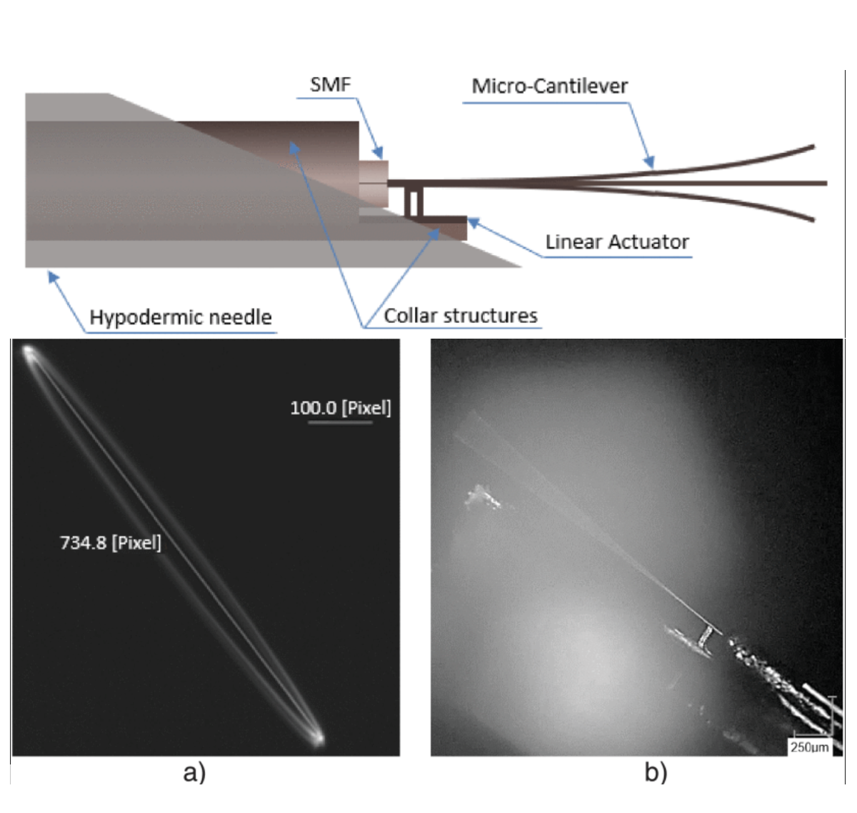
Kaur, Mandeep; Brown, Malcolm; Lane, Pierre M.; Menon, Carlo
An Electro-Thermally Actuated Micro-Cantilever-Based Fiber Optic Scanner Journal Article
In: IEEE Sensors Journal , vol. 20, iss. 17, pp. 9877-9885, 2020.
@article{Kaur2020,

title = {An Electro-Thermally Actuated Micro-Cantilever-Based Fiber Optic Scanner},

author = {Mandeep Kaur and Malcolm Brown and Pierre M. Lane and Carlo Menon},

url = {https://doi.org/10.1109/JSEN.2020.2992371, DOI},

year  = {2020},

date = {2020-09-01},

urldate = {2020-09-01},

journal = {IEEE Sensors Journal },

volume = {20},

issue = {17},

pages = {9877-9885},

abstract = {A sub-millimeter sized optical scanner driven by electro-thermal actuation is presented. The scanner is composed of a single-mode optical fiber (SMF) with a cantilevered section at its distal tip. The fiber cantilever is electrothermally actuated near its base in a single direction and excited at resonance to obtain large deflectionsat the tip of the fiber. Two-dimensional imaging of an object is demonstrated by simultaneously rotating the object while scanning across its diameter. Illumination light from the optical core of the fiber cantilever is projected through a lens onto the object. Reflected light is collected by the same lens and projected onto a photodetector. An image of the object is reconstructed by interpolation of the detected signal. The resolution of the system was measured to be 16μm by imaging a resolution target. The electro-thermal fiber actuator may provide a new technique for scanning in sub-millimeter sized forward-viewing endoscopic catheters.},

keywords = {},

pubstate = {published},

tppubtype = {article}

}
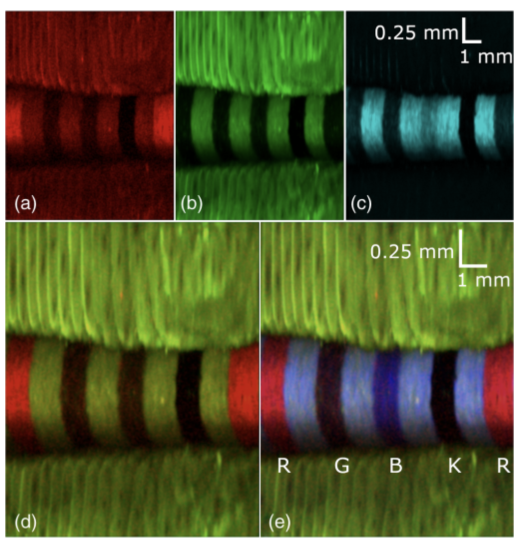
Buenconsejo, Andrea Louise; Hohert, Geoffrey; Manning, Max; Abouei, Elham; Tingley, Reid; Janzen, Ian; McAlpine, Jessica; Miller, Dianne; Lee, Anthony; Lane, Pierre; MacAulay, Calum
In: Journal of Biomedical Optics , vol. 25, iss. 3, no. 032005, 2019.
@article{Buenconsejo2019,

title = {Submillimeter diameter rotary-pullback fiber-optic endoscope for narrowband red-green-blue reflectance, optical coherence tomography, and autofluorescence in vivo imaging},

author = {Andrea Louise Buenconsejo and Geoffrey Hohert and Max Manning and Elham Abouei and Reid Tingley and Ian Janzen and Jessica McAlpine and Dianne Miller and Anthony Lee and Pierre Lane and Calum MacAulay},

url = {https://doi.org/10.1117/1.JBO.25.3.032005, DOI

https://biophotonics.bccrc.ca/wp-content/uploads/2022/02/032005_1.pdf, Full Text (PDF)},

year  = {2019},

date = {2019-10-24},

urldate = {2019-10-24},

journal = {Journal of Biomedical Optics },

volume = {25},

number = {032005},

issue = {3},

abstract = {A fiber-based endoscopic imaging system combining narrowband red-green-blue (RGB) reflectance with optical coherence tomography (OCT) and autofluorescence imaging (AFI) has been developed. The system uses a submillimeter diameter rotary-pullback double-clad fiber imaging catheter for sample illumination and detection. The imaging capabilities of each modality are presented and demonstrated with images of a multicolored card, fingerprints, and tongue mucosa. Broadband imaging, which was done to compare with narrowband sources, revealed better contrast but worse color consistency compared with narrowband RGB reflectance. The measured resolution of the endoscopic system is 25  μm in both the rotary direction and the pullback direction. OCT can be performed simultaneously with either narrowband RGB reflectance imaging or AFI.},

keywords = {},

pubstate = {published},

tppubtype = {article}

}
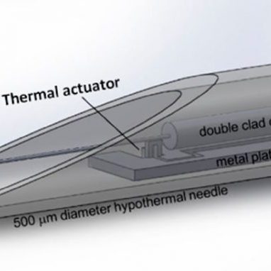
Ahrabi, Aydin Aghajanzadeh; Kaur, Mandeep; Li, Yasong; Lane, Pierre; Menon, Carlo
An Electro-Thermal Actuation Method for Resonance Vibration of a Miniaturized Optical-Fiber Scanner for Future Scanning Fiber Endoscope Design Journal Article
In: Actuators, vol. 8, no. 1, 2019.
@article{Ahrabi2019,

title = {An Electro-Thermal Actuation Method for Resonance Vibration of a Miniaturized Optical-Fiber Scanner for Future Scanning Fiber Endoscope Design},

author = {Aydin Aghajanzadeh Ahrabi and Mandeep Kaur and Yasong Li and Pierre Lane and Carlo Menon},

url = {https://biophotonics.bccrc.ca/wp-content/uploads/2019/03/Ahrabi-2019.pdf, Full Text (PDF)

https://doi.org/10.3390/act8010021, DOI},

year  = {2019},

date = {2019-03-01},

journal = {Actuators},

volume = {8},

number = {1},

abstract = {Medical professionals increasingly rely on endoscopes to carry out many minimally invasive procedures on patients to safely examine, diagnose, and treat a large variety of conditions. However, their insertion tube diameter dictates which passages of the body they can be inserted into and, consequently, what organs they can access. For inaccessible areas and organs, patients often undergo invasive and risky procedures—diagnostic confirmation of peripheral lung nodules via transthoracic needle biopsy is one example from oncology. Hence, this work sets out to present an optical-fiber scanner for a scanning fiber endoscope design that has an insertion tube diameter of about 0.5 mm, small enough to be inserted into the smallest airways of the lung. To attain this goal, a novel approach based on resonance thermal excitation of a single-mode 0.01-mm-diameter fiber-optic cantilever oscillating at 2–4 kHz is proposed. The small size of the electro-thermal actuator enables miniaturization of the insertion tube. Lateral free-end deflection of the cantilever is used as a benchmark for evaluating performance. Experimental results show that the cantilever can achieve over 0.2 mm of displacement at its free end. The experimental results also support finite element simulation models which can be used for future design iterations of the endoscope.},

keywords = {},

pubstate = {published},

tppubtype = {article}

}
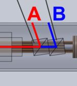
Lee, Anthony M. D.; MacAulay, Calum; Lane, Pierre
Depth-multiplexed optical coherence tomography dual-beam manually-actuated distortion-corrected imaging (DMDI) with a micromotor catheter Journal Article
In: Optics Express, vol. 9, no. 11, pp. 5678-5690, 2018.
@article{Lee2018,

title = {Depth-multiplexed optical coherence  tomography dual-beam manually-actuated  distortion-corrected imaging (DMDI) with a  micromotor catheter},

author = { Anthony M.D. Lee and Calum MacAulay and Pierre Lane},

url = {https://biophotonics.bccrc.ca/wp-content/uploads/2018/10/Lee-2018.pdf, Full Text (PDF)

https://doi.org/10.1364/BOE.9.005678, DOI},

year  = {2018},

date = {2018-11-01},

journal = {Optics Express},

volume = {9},

number = {11},

pages = {5678-5690},

abstract = {We present a new micromotor catheter implementation of dual-beam manuallyactuated distortion-corrected imaging (DMDI). The new catheter, called a depth-multiplexed dual-beam micromotor catheter, or mDBMC, maintains the primary advantage of unlimited field-of-view distortion-corrected imaging along the catheter axis. The mDBMC uses a polarization beam splitter and cube mirror to create two beams that scan circularly with approximately constant separation at the catheter surface. This arrangement also multiplexes both imaging channels into a single optical coherence tomography channel by offsetting them in depth, requiring half the data bandwidth compared to previous DMDI demonstrations that used two parallel image acquisition systems. Furthermore, the relatively simple scanning pattern of the two beams enables a straightforward automated distortion correction algorithm. We demonstrate the imaging capabilities of this catheter with a printed paper phantom and in a section of dragon fruit.},

keywords = {},

pubstate = {published},

tppubtype = {article}

}
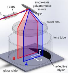
Harlow, Madeline; MacAulay, Calum; Lane, Pierre; Lee, Anthony M. D.
Dual-beam manually actuated distortion corrected imaging (DMDI): two dimensional scanning with a single-axis galvanometer Journal Article
In: Optics Express, vol. 26, no. 14, pp. 18758-18772, 2018.
@article{Harlow2018,

title = {Dual-beam manually actuated distortion corrected imaging (DMDI): two dimensional scanning with a single-axis galvanometer},

author = {Madeline Harlow and Calum MacAulay and Pierre Lane and Anthony M.D. Lee

},

url = {https://biophotonics.bccrc.ca/wp-content/uploads/2018/10/Harlow-2018.pdf, Full Text (PDF)},

doi = {10.1364/OE.26.018758},

year  = {2018},

date = {2018-07-09},

journal = {Optics Express},

volume = {26},

number = {14},

pages = {18758-18772},

abstract = {We recently demonstrated a new two-dimensional imaging paradigm called dual-beam manually actuated distortion-corrected imaging (DMDI). This technique uses a single mechanical scanner and two spatially separated beams to determine relative sample velocity and simultaneously corrects image distortions due to manual actuation. DMDI was first demonstrated using a rotating dual-beam micromotor catheter. Here, we present a new

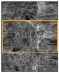
implementation of DMDI using a single axis galvanometer to scan a pair of beams in approximately parallel lines onto a sample. Furthermore, we present a method for automated distortion correction based on frame co-registration between images acquired by the two beams. Distortion correction is possible for manually actuated motion both perpendicular and parallel to the galvanometer-scanned lines. Using en face OCT as the imaging modality, we demonstrate DMDI and the automated distortion correction algorithm for imaging a printed

paper phantom, a dragon fruit, and a fingerprint.},

keywords = {},

pubstate = {published},

tppubtype = {article}

}
implementation of DMDI using a single axis galvanometer to scan a pair of beams in approximately parallel lines onto a sample. Furthermore, we present a method for automated distortion correction based on frame co-registration between images acquired by the two beams. Distortion correction is possible for manually actuated motion both perpendicular and parallel to the galvanometer-scanned lines. Using en face OCT as the imaging modality, we demonstrate DMDI and the automated distortion correction algorithm for imaging a printed
paper phantom, a dragon fruit, and a fingerprint.
Abouei, Elham; Lee, Anthony M. D.; Pahlevaninezhad, Hamid; Hohert, Geoffrey; Cua, Michelle; Lane, Pierre; Lam, Stephen; MacAulay, Calum
In: Journal of Biomedical Optics, vol. 23, no. 1, pp. 016004, 2018.
@article{Abouei2018,

title = {Correction of motion artifacts in endoscopic optical coherence tomography and autofluorescence images based on azimuthal en face image registration},

author = {Elham Abouei and Anthony M. D. Lee and Hamid Pahlevaninezhad and Geoffrey Hohert and Michelle Cua and Pierre Lane and Stephen Lam and Calum MacAulay},

url = {https://biophotonics.bccrc.ca/pubs/Abouei-2018.pdf, Full Text (PDF)},

doi = {10.1117/1.JBO.23.1.016004},

year  = {2018},

date = {2018-01-04},

journal = {Journal of Biomedical Optics},

volume = {23},

number = {1},

pages = {016004},

abstract = {We present a method for the correction of motion artifacts present in two- and three-dimensional in vivo endoscopic images produced by rotary-pullback catheters. This method can correct for cardiac / breathing based motion artifacts and catheter-based motion artifacts such as nonuniform rotational distortion (NURD). This method assumes that en face tissue imaging contains slowly varying structures that are roughly parallel to the pullback axis. The method reduces motion artifacts using a dynamic time warping solution through a cost matrix that measures similarities between adjacent frames in en face images. We optimize and demonstrate the suitability of this method using a real and simulated NURD phantom and in vivo endoscopic pulmonary optical coherence tomography and autofluorescence images. Qualitative and quantitative evaluations of the method show an enhancement of the image quality.},

keywords = {},

pubstate = {published},

tppubtype = {article}

}
Hill, Breana; Lam, Sylvia F; Lane, Pierre; MacAulay, Calum; Fradkin, Leonid; Follen, Michele
Established and Emerging Optical Technologies for the Real-Time Detection of Cervical Neoplasia: A Review Journal Article
In: Journal of Cancer Therapy, vol. 8, pp. 1241-1278, 2017.
@article{Hill12017,

title = {Established and Emerging Optical Technologies for the Real-Time Detection of Cervical Neoplasia: A Review},

author = {Breana Hill and Sylvia F Lam and Pierre Lane and Calum MacAulay and Leonid Fradkin and

Michele Follen},

url = {https://biophotonics.bccrc.ca/pubs/Hill-2017.pdf, Full Text (PDF)},

doi = {10.4236/jct.2017.813105},

year  = {2017},

date = {2017-12-21},

journal = {Journal of Cancer Therapy},

volume = {8},

pages = {1241-1278},

abstract = {Cervical cancer remains a critically important problem for women, especially those women in the developing world where the case-fatality rate is high. There are an estimated 528,000 cases and 266,000 deaths worldwide. Established screening and detection programs in the developed world have lowered the mortality from 40/100,000 to 2/100,000 over the last 60 years. The standard of care has been and continues to be: a screening Papanicolaou smear with or without Human Papilloma Virus (HPV) testing; followed by colposcopy and biopsies and if the smear is abnormal; and followed by treatment if the biopsies show high grade disease (cervical intraepithelial neoplasia (CIN) grades 2 and 3 and Carcinoma-in-situ). Low grade lesions (Pap smears with Atypical Cells of Uncertain Significance (ASCUS), Low Grade Squamous Intraepithelial Lesions (LGSIL), biopsies showing HPV changes or showing CIN 1); are usually followed for two years and then treated if persistent. Treatment can be performed with loop excision, LASER, or cryotherapy. Loop excision yields a specimen which can be reviewed to establish the diagnosis more accurately. LASER vaporizes the lesion and cryotherapy leads to tissue destruction. Under long term study; loop excision, LASER, and cryotherapy have the same rate of cure. The standard of care is expensive and takes 6 - 12 weeks for the individual patient. During the last twenty years, new technologies that can view the cervix and even image the cervix with cellular resolution have been developed. These technologies could lead to a new paradigm in which diagnosis and treatment occurs at a single visit. These technologies include fluorescence and reflectance spectroscopy (probe or wide-field, whole cervix scanning approaches) and fluorescence confocal endomicroscopy or high resolution micro-endoscopy. Both technologies have received Federal Drug Administration (FDA) and have been commercialized. Research trials continue to show their remarkable performance. These technologies are reviewed and clinical trials are summarized. Emerging technologies are coming along that may compete with those already approved and include optical coherence tomography, optical coherence tomography with autofluorescence, diffuse optical microscopy, and dual mode micro-endoscopy. These technologies are also reviewed and where available, clinical data is reported. Optical technologies are ready to diffuse into clinical practice because they will save money and 3 or 4 visits in the developed world and offer the same standard of care to the developing world where more cervical cancer exists. },

keywords = {},

pubstate = {published},

tppubtype = {article}

}
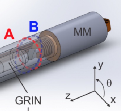
Lee, Anthony M. D.; Hohert, Geoffrey; Angkiriwang, Patricia T.; MacAulay, Calum; Lane, Pierre
Dual-beam manually-actuated distortion-corrected imaging (DMDI) with micromotor catheters Journal Article
In: Optics Express, vol. 25, no. 18, pp. 22164-22177, 2017.
@article{Lee2017,

title = {Dual-beam manually-actuated distortion-corrected imaging (DMDI) with micromotor catheters},

author = {Anthony M. D. Lee and Geoffrey Hohert and Patricia T. Angkiriwang and Calum MacAulay and Pierre Lane},

url = {https://biophotonics.bccrc.ca/pubs/Lee-2017.pdf, Full Text (PDF)},

doi = {10.1364/OE.25.022164},

year  = {2017},

date = {2017-09-01},

journal = {Optics Express},

volume = {25},

number = {18},

pages = {22164-22177},

abstract = {We present a new paradigm for performing two-dimensional scanning called dual-beam manually-actuated distortion-corrected imaging (DMDI). DMDI operates by imaging the same object with two spatially-separated beams that are being mechanically scanned rapidly in one dimension with slower manual actuation along a second dimension. Registration of common features between the two imaging channels allows remapping of the images to correct for distortions due to manual actuation. We demonstrate DMDI using a 4.7 mm OD rotationally scanning dual-beam micromotor catheter (DBMC). The DBMC requires a simple, one-time calibration of the beam paths by imaging a patterned phantom. DMDI allows for distortion correction of non-uniform axial speed and rotational motion of the DBMC. We show the utility of this technique by demonstrating en face OCT image distortion correction of a manually-scanned checkerboard phantom and fingerprint scan.},

keywords = {},

pubstate = {published},

tppubtype = {article}

}
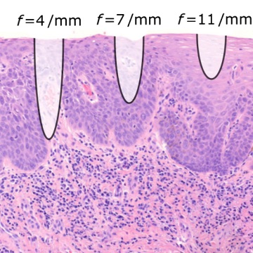
Bodenschatz, Nico; Poh, Catherine F.; Lam, Sylvia; Lane, Pierre M.; Guillaud, Martial; MacAulay, Calum E.
Dual-mode endomicroscopy for detection of epithelial dysplasia in the mouth: a descriptive pilot study Journal Article
In: Journal of Biomedical Optics, vol. 22, no. 8, pp. 086005, 2017.
@article{Bodenschatz2017,

title = {Dual-mode endomicroscopy for detection of epithelial dysplasia in the mouth: a descriptive pilot study},

author = {Nico Bodenschatz and Catherine F. Poh and Sylvia Lam and Pierre M. Lane and Martial Guillaud and Calum E. MacAulay},

url = {https://biophotonics.bccrc.ca/pubs/Bodenschatz-2017.pdf, Full Text (PDF)},

doi = {10.1117/1.JBO.22.8.086005},

year  = {2017},

date = {2017-08-19},

journal = {Journal of Biomedical Optics},

volume = {22},

number = {8},

pages = {086005},

abstract = {Dual-mode endomicroscopy is a diagnostic tool for early cancer detection. It combines the high-resolution nuclear tissue contrast of fluorescence endomicroscopy with quantified depth-dependent epithelial backscattering as obtained by diffuse optical microscopy. In an in vivo pilot imaging study of 27 oral lesions from 21 patients, we demonstrate the complementary diagnostic value of both modalities and show correlations between grade of epithelial dysplasia and relative depth-dependent shifts in light backscattering. When combined, the two modalities provide diagnostic sensitivity to both moderate and severe epithelial dysplasia in vivo.},

keywords = {},

pubstate = {published},

tppubtype = {article}

}
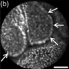
Schlosser, Colin; Bodenschatz, Nico; Lam, Sylvia; Lee, Marette; McAlpine, Jessica N.; Miller, Dianne M.; Niekerk, Dirk J. T. Van; Follen, Michele; Guillaud, Martial; MacAulay, Calum E.; Lane, Pierre M.
Fluorescence confocal endomicroscopy of the cervix: pilot study on the potential and limitations for clinical implementation Journal Article
In: Journal of Biomedical Optics, vol. 21, no. 12, pp. 126011, 2016.
@article{Schlosser2016,

title = {Fluorescence confocal endomicroscopy of the cervix: pilot study on the potential and limitations for clinical implementation},

author = {Colin Schlosser and Nico Bodenschatz and Sylvia Lam and Marette Lee and Jessica N. McAlpine and Dianne M. Miller and Dirk J. T. Van Niekerk and Michele Follen and Martial Guillaud and Calum E. MacAulay and Pierre M. Lane},

url = {https://biophotonics.bccrc.ca/pubs/Schlosser-2016.pdf, Full Text (PDF)},

doi = {10.1117/1. JBO.21.12.126011},

year  = {2016},

date = {2016-12-01},

journal = {Journal of Biomedical Optics},

volume = {21},

number = {12},

pages = {126011},

abstract = {Current diagnostic capabilities and limitations of fluorescence endomicroscopy in the cervix are assessed by qualitative and quantitative image analysis. Four cervical tissue types are investigated: normal columnar epithelium, normal and precancerous squamous epithelium, and stromal tissue. This study focuses on the perceived variability within and the subtle differences between the four tissue groups in the context of endomicroscopic in vivo pathology. Conclusions are drawn on the general ability to distinguish and diagnose tissue types, on the need for imaging depth control to enhance differentiation, and on the possible risks for diagnostic misinterpretations.},

keywords = {},

pubstate = {published},

tppubtype = {article}

}
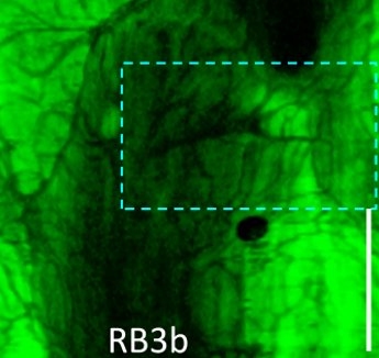
Pahlevaninezhad, Hamid; Lee, Anthony MD; Hohert, Geoffrey; Lam, Stephen; Shaipanich, Tawimas; Beaudoin, Eve-Lea; MacAulay, Calum; Boudoux, Caroline; Lane, Pierre
Endoscopic high-resolution autofluorescence imaging and OCT of pulmonary vascular networks Journal Article
In: Optics Letters, vol. 41, no. 14, pp. 3209-32-12, 2016.
@article{Pahlevaninezhad2016,

title = {Endoscopic high-resolution autofluorescence imaging and OCT of pulmonary vascular networks},

author = {Hamid Pahlevaninezhad and Anthony MD Lee and Geoffrey Hohert and Stephen Lam and Tawimas Shaipanich and Eve-Lea Beaudoin and Calum MacAulay and Caroline Boudoux and Pierre Lane},

url = {https://biophotonics.bccrc.ca/pubs/Pahlevaninezhad-2016.pdf, Full Text (PDF)},

doi = {10.1364/OL.41.003209},

year  = {2016},

date = {2016-07-15},

journal = {Optics Letters},

volume = {41},

number = {14},

pages = {3209-32-12},

abstract = {High-resolution imaging from within airways may allow new methods for studying lung disease. In this work, we report an endoscopic imaging system capable of high-resolution autofluorescence imaging (AFI) and optical coherence tomography (OCT) in peripheral airways using a 0.9 mm diameter double-clad fiber (DCF) catheter. In this system, AFI excitation light is coupled into the core of the DCF, enabling tightly focused excitation light while maintaining efficient collection of autofluorescence emission through the large diameter inner cladding of the DCF. We demonstrate the ability of this imaging system to visualize pulmonary vasculature as small as 12 μm in vivo.},

keywords = {},

pubstate = {published},

tppubtype = {article}

}
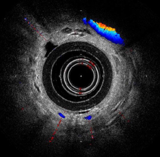
Kirby, Miranda; Lane, Pierre; Coxson, Harvey
Measurement of pulmonary structure and function Book Chapter
In: Barr, R. Graham; Parr, David G.; Vogel-Claussen, Jens (Ed.): Imaging, no. 70, Chapter 14, pp. 216-232, European Respiratory Society, 2016.
@inbook{Kirby2016,

title = {Measurement of pulmonary structure and function},

author = {Miranda Kirby and Pierre Lane and Harvey Coxson},

editor = {R. Graham Barr and David G. Parr and Jens Vogel-Claussen},

doi = {10.1183/2312508X.10003415},

year  = {2016},

date = {2016-03-01},

booktitle = {Imaging},

number = {70},

pages = {216-232},

publisher = {European Respiratory Society},

chapter = {14},

abstract = {Much of our understanding of both healthy and diseased lung is based on quantitative morphological assessment using pathology.  However, the development of new therapies for lung disease requires measurements and tools that can non-invasively measure lung structure and function both on a global and a regional scale.  Medical imaging technologies allows us to “see” the complex internal structures of the lung in three-dimensions so that detailed anatomic measurements can be performed, while other imaging modalities can also measure how the lung functions.  Here we have highlighted three techniques that have impacted pulmonary imaging research with emphasis on quantitative measurements:  computed tomography (CT), hyperpolarized noble gas magnetic resonance imaging (MRI) and optical coherence tomography (OCT).  For each of these imaging modalities, we discuss the quantitative measurements that can be derived as well as discuss the pulmonary diseases where each technique has demonstrated utility, discuss some drawbacks that are unique to each technique, and, finally, discuss some important directions for future research.},

keywords = {},

pubstate = {published},

tppubtype = {inbook}

}
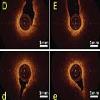
Kirby, Miranda; Ohtani, Keishi; Nickens, Taylor; Lisbona, Rosa Maria Lopez; Lee, Anthony M. D.; Shaipanich, Tawimas; Lane, Pierre; MacAulay, Calum; Lam, Stephen; Coxson, Harvey O.
Reproducibility of optical coherence tomography airway imaging Journal Article
In: Biomed. Opt. Express, vol. 6, no. 11, pp. 4365–4377, 2015.
@article{Kirby:15,

title = {Reproducibility of optical coherence tomography airway imaging},

author = {Miranda Kirby and Keishi Ohtani and Taylor Nickens and Rosa Maria Lopez Lisbona and Anthony M. D. Lee and Tawimas Shaipanich and Pierre Lane and Calum MacAulay and Stephen Lam and Harvey O. Coxson},

url = {https://biophotonics.bccrc.ca/pubs/Kirby-2015a.pdf, Full Text (PDF)

},

doi = {10.1364/BOE.6.004365},

year  = {2015},

date = {2015-11-01},

journal = {Biomed. Opt. Express},

volume = {6},

number = {11},

pages = {4365--4377},

publisher = {OSA},

abstract = {Optical coherence tomography (OCT) is a promising imaging technique to evaluate small airway remodeling. However, the short-term insertion-reinsertion reproducibility of OCT for evaluating the same bronchial pathway has yet to be established. We evaluated 74 OCT data sets from 38 current or former smokers twice within a single imaging session. Although the overall insertion-reinsertion airway wall thickness (WT) measurement coefficient of variation (CV) was moderate at 12%, much of the variability between repeat imaging was attributed to the observer; CV for repeated measurements of the same airway (intra-observer CV) was 9%. Therefore, reproducibility may be improved by introduction of automated analysis approaches suggesting that OCT has potential to be an in-vivo method for evaluating airway remodeling in future longitudinal and intervention studies.},

keywords = {},

pubstate = {published},

tppubtype = {article}

}
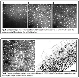
Sheikhzadeh, Fahime; Ward, Rabab K.; Carraro, Anita; Chen, Zhao Yang; van Niekerk, Dirk; Miller, Dianne; Ehlen, Tom; MacAulay, Calum E.; Follen, Michele; Lane, Pierre M.; Guillaud, Martial
Quantification of confocal fluorescence microscopy for the detection of cervical intraepithelial neoplasia Journal Article
In: Biomedical Engineering Online, vol. 14, no. 96, 2015.
@article{Sheikhzadeh2015,

title = {Quantification of confocal fluorescence microscopy for the detection of cervical intraepithelial neoplasia},

author = {Fahime Sheikhzadeh and Rabab K. Ward and Anita Carraro and Zhao Yang Chen and Dirk van Niekerk and Dianne Miller and Tom Ehlen and Calum E. MacAulay and Michele Follen and Pierre M. Lane and Martial Guillaud },

url = {https://biophotonics.bccrc.ca/pubs/Sheikhzadeh-2015.pdf, Full Text (PDF)},

doi = {10.1186/s12938-015-0093-6},

year  = {2015},

date = {2015-10-24},

journal = {Biomedical Engineering Online},

volume = {14},

number = {96},

abstract = {Background

Cervical cancer remains a major health problem, especially in developing countries. Colposcopic examination is used to detect high-grade lesions in patients with a history of abnormal pap smears. New technologies are needed to improve the sensitivity and specificity of this technique. We propose to test the potential of fluorescence confocal microscopy to identify high-grade lesions.


Methods

We examined the quantification of ex vivo confocal fluorescence microscopy to differentiate among normal cervical tissue, low-grade Cervical Intraepithelial Neoplasia (CIN), and high-grade CIN. We sought to (1) quantify nuclear morphology and tissue architecture features by analyzing images of cervical biopsies; and (2) determine the accuracy of high-grade CIN detection via confocal microscopy relative to the accuracy of detection by colposcopic impression. Forty-six biopsies obtained from colposcopically normal and abnormal cervical sites were evaluated. Confocal images were acquired at different depths from the epithelial surface and histological images were analyzed using in-house software.


Results

The features calculated from the confocal images compared well with those features obtained from the histological images and histopathological reviews of the specimens (obtained by a gynecologic pathologist). The correlations between two of these features (the nuclear-cytoplasmic ratio and the average of three nearest Delaunay-neighbors distance) and the grade of dysplasia were higher than that of colposcopic impression. The sensitivity of detecting high-grade dysplasia by analysing images collected at the surface of the epithelium, and at 15 and 30 μm below the epithelial surface were respectively 100, 100, and 92 %.


Conclusions

Quantitative analysis of confocal fluorescence images showed its capacity for discriminating high-grade CIN lesions vs. low-grade CIN lesions and normal tissues, at different depth of imaging. This approach could be used to help clinicians identify high-grade CIN in clinical settings.},

keywords = {},

pubstate = {published},

tppubtype = {article}

}
Cervical cancer remains a major health problem, especially in developing countries. Colposcopic examination is used to detect high-grade lesions in patients with a history of abnormal pap smears. New technologies are needed to improve the sensitivity and specificity of this technique. We propose to test the potential of fluorescence confocal microscopy to identify high-grade lesions.
Methods
We examined the quantification of ex vivo confocal fluorescence microscopy to differentiate among normal cervical tissue, low-grade Cervical Intraepithelial Neoplasia (CIN), and high-grade CIN. We sought to (1) quantify nuclear morphology and tissue architecture features by analyzing images of cervical biopsies; and (2) determine the accuracy of high-grade CIN detection via confocal microscopy relative to the accuracy of detection by colposcopic impression. Forty-six biopsies obtained from colposcopically normal and abnormal cervical sites were evaluated. Confocal images were acquired at different depths from the epithelial surface and histological images were analyzed using in-house software.
Results
The features calculated from the confocal images compared well with those features obtained from the histological images and histopathological reviews of the specimens (obtained by a gynecologic pathologist). The correlations between two of these features (the nuclear-cytoplasmic ratio and the average of three nearest Delaunay-neighbors distance) and the grade of dysplasia were higher than that of colposcopic impression. The sensitivity of detecting high-grade dysplasia by analysing images collected at the surface of the epithelium, and at 15 and 30 μm below the epithelial surface were respectively 100, 100, and 92 %.
Conclusions
Quantitative analysis of confocal fluorescence images showed its capacity for discriminating high-grade CIN lesions vs. low-grade CIN lesions and normal tissues, at different depth of imaging. This approach could be used to help clinicians identify high-grade CIN in clinical settings.

Pahlevaninezhad, Hamid; Lee, Anthony M. D.; Ritchie, Alexander; Shaipanich, Tawimas; Zhang, Wei; Ionescu, Diana N.; Hohert, Geoffrey; MacAulay, Calum; Lam, Stephen; Lane, Pierre
Endoscopic Doppler optical coherence tomography and autofluorescence imaging of peripheral pulmonary nodules and vasculature Journal Article
In: Biomedical Optics Express, vol. 6, no. 10, pp. 4191-4199, 2015.
@article{Pahlevaninezhad:15,

title = {Endoscopic Doppler optical coherence tomography and autofluorescence imaging of peripheral pulmonary nodules and vasculature},

author = {Hamid Pahlevaninezhad and Anthony M. D. Lee and Alexander Ritchie and Tawimas Shaipanich and Wei Zhang and Diana N. Ionescu and Geoffrey Hohert and Calum MacAulay and Stephen Lam and Pierre Lane},

url = {https://biophotonics.bccrc.ca/pubs/Pahlevaninezhad-2015.pdf, Full Text (PDF)},

doi = {10.1364/BOE.6.004191},

year  = {2015},

date = {2015-10-01},

journal = {Biomedical Optics Express},

volume = {6},

number = {10},

pages = {4191-4199},

publisher = {OSA},

abstract = {We present the first endoscopic Doppler optical coherence tomography and co-registered autofluorescence imaging (DOCT-AFI) of peripheral pulmonary nodules and vascular networks in vivo using a small 0.9 mm diameter catheter. Using exemplary images from volumetric data sets collected from 31 patients during flexible bronchoscopy, we demonstrate how DOCT and AFI offer complementary information that may increase the ability to locate and characterize pulmonary nodules. AFI offers a sensitive visual presentation for the rapid identification of suspicious airway sites, while co-registered OCT provides detailed structural information to assess the airway morphology. We demonstrate the ability of AFI to visualize vascular networks in vivo and validate this finding using Doppler and structural OCT. Given the advantages of higher resolution, smaller probe size, and ability to visualize vasculature, DOCT-AFI has the potential to increase diagnostic accuracy and minimize bleeding to guide biopsy of pulmonary nodules compared to radial endobronchial ultrasound, the current standard of care.},

keywords = {},

pubstate = {published},

tppubtype = {article}

}
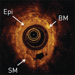
Kirby, Miranda; Ohtani, Keishi; Lisbona, Rosa Maria Lopez; Lee, Anthony MD; Zhang, Wei; Lane, Pierre; Varfolomeva, Nina; Hui, Linda; Ionescu, Diana; Coxson, Harvey O; MacAulay, Calum; FitzGerald, J. Mark; Lam, Stephen
Bronchial thermoplasty in asthma: 2-year follow-up using optical coherence tomography Journal Article
In: European Respiratory Journal, vol. 46, no. 3, pp. 859-862, 2015.
@article{kirby2015bronchial,

title = {Bronchial thermoplasty in asthma: 2-year follow-up using optical coherence tomography},

author = { Miranda Kirby and Keishi Ohtani and Rosa Maria Lopez Lisbona and Anthony MD Lee and Wei Zhang and Pierre Lane and Nina Varfolomeva and Linda Hui and Diana Ionescu and Harvey O Coxson and Calum MacAulay and J. Mark FitzGerald and Stephen Lam},

url = {https://biophotonics.bccrc.ca/pubs/Kirby-2015.pdf, Full Text (PDF)},

doi = {10.1183/09031936.00016815},

year  = {2015},

date = {2015-01-01},

journal = {European Respiratory Journal},

volume = {46},

number = {3},

pages = {859-862},

publisher = {Eur Respiratory Soc},

abstract = {We evaluated two severe asthmatics immediately prior to and longitudinally following BT, and demonstrated a reduction in airway wall thickness that persisted 2 years following treatment in the BT responder, as well as differences in airway wall features between the responder and nonresponder prior to treatment. These observations generate hypotheses for a larger study to determine if airway changes defined by OCT imaging can identify asthma patients who will benefit from BT and to determine the long-term effects of the treatment.},

keywords = {},

pubstate = {published},

tppubtype = {article}

}
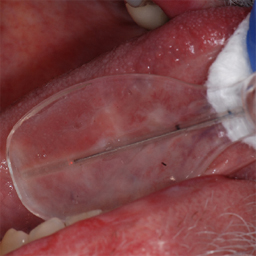
Lee, Anthony MD; Cahill, Lucas; Liu, Kelly; MacAulay, Calum; Poh, Catherine; Lane, Pierre
Wide-field in vivo oral OCT imaging Journal Article
In: Biomedical Optics Express, vol. 6, no. 7, pp. 2664-2674, 2015.
@article{lee2015wide,

title = {Wide-field in vivo oral OCT imaging},

author = { Anthony MD Lee and Lucas Cahill and Kelly Liu and Calum MacAulay and Catherine Poh and Pierre Lane},

url = {https://biophotonics.bccrc.ca/pubs/Lee-2015.pdf, Full Text (PDF)

http://www.ncbi.nlm.nih.gov/pubmed/26203389, PubMed},

doi = {10.1364/BOE.6.002664},

year  = {2015},

date = {2015-01-01},

journal = {Biomedical Optics Express},

volume = {6},

number = {7},

pages = {2664-2674},

publisher = {Optical Society of America},

abstract = {We have built a polarization-sensitive swept source Optical Coherence Tomography (OCT) instrument capable of wide-field in vivo imaging in the oral cavity. This instrument uses a hand-held side-looking fiber-optic rotary pullback catheter that can cover two dimensional tissue imaging fields approximately 2.5 mm wide by up to 90 mm length in a single image acquisition. The catheter spins at 100 Hz with pullback speeds up to 15 mm/s allowing imaging of areas up to 225 mm2 field-of-view in seconds. A catheter sheath and two optional catheter sheath holders have been designed to allow imaging at all locations within the oral cavity. Image quality of 2-dimensional image slices through the data can be greatly enhanced by averaging over the orthogonal dimension to reduce speckle. Initial in vivo imaging results reveal a wide-field view of features such as epithelial thickness and continuity of the basement membrane that may be useful in clinic for chair-side management of oral lesions.},

keywords = {},

pubstate = {published},

tppubtype = {article}

}
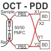
Lee, Anthony; Pahlevaninezhad, Hamid; Yang, Victor XD; Lam, Stephen; MacAulay, Calum; Lane, Pierre
Fiber-optic polarization diversity detection for rotary probe optical coherence tomography Journal Article
In: Optics Letters, vol. 39, no. 12, pp. 3638–3641, 2014.
@article{lee2014fiber,

title = {Fiber-optic polarization diversity detection for rotary probe optical coherence tomography},

author = { Anthony Lee and Hamid Pahlevaninezhad and Victor XD Yang and Stephen Lam and Calum MacAulay and Pierre Lane},

url = {https://biophotonics.bccrc.ca/pubs/Lee-2014.pdf, Full Text (PDF)

http://www.ncbi.nlm.nih.gov/pubmed/24978556, Pubmed},

doi = {10.1364/OL.39.003638},

year  = {2014},

date = {2014-01-01},

journal = {Optics Letters},

volume = {39},

number = {12},

pages = {3638--3641},

publisher = {Optical Society of America},

abstract = {We report a polarization diversity detection scheme for optical coherence tomography with a new, custom, miniaturized fiber coupler with single mode (SM) fiber inputs and polarization maintaining (PM) fiber outputs. The SM fiber inputs obviate matching the optical lengths of the X and Y OCT polarization channels prior to interference and the PM fiber outputs ensure defined X and Y axes after interference. Advantages for this scheme include easier alignment, lower cost, and easier miniaturization compared to designs with free-space bulk optical components. We demonstrate the utility of the detection system to mitigate the effects of rapidly changing polarization states when imaging with rotating fiber optic probes in Intralipid suspension and during in vivo imaging of human airways.},

keywords = {},

pubstate = {published},

tppubtype = {article}

}
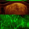
Pahlevaninezhad, Hamid; Lee, Anthony; Shaipanich, Tawimas; Raizada, Rashika; Cahill, Lucas; Hohert, Geoffrey; Yang, Victor XD; Lam, Stephen; MacAulay, Calum; Lane, Pierre
A high-efficiency fiber-based imaging system for co-registered autofluorescence and optical coherence tomography Journal Article
In: Biomedical optics express, vol. 5, no. 9, pp. 2978–2987, 2014.
@article{pahlevaninezhad2014high,

title = {A high-efficiency fiber-based imaging system for co-registered autofluorescence and optical coherence tomography},

author = { Hamid Pahlevaninezhad and Anthony Lee and Tawimas Shaipanich and Rashika Raizada and Lucas Cahill and Geoffrey Hohert and Victor XD Yang and Stephen Lam and Calum MacAulay and Pierre Lane},

url = {https://biophotonics.bccrc.ca/pubs/Pahlevaninezhad-2014.pdf, Full Text (PDF)

http://www.ncbi.nlm.nih.gov/pubmed/25401011, PubMed},

doi = {10.1364/BOE.5.002978},

year  = {2014},

date = {2014-01-01},

journal = {Biomedical optics express},

volume = {5},

number = {9},

pages = {2978--2987},

publisher = {Optical Society of America},

abstract = {We present a power-efficient fiber-based imaging system capable of co-registered autofluorescence imaging and optical coherence tomography (AF/OCT). The system employs a custom fiber optic rotary joint (FORJ) with an embedded dichroic mirror to efficiently combine the OCT and AF pathways. This three-port wavelength multiplexing FORJ setup has a throughput of more than 83% for collected AF emission, significantly more efficient compared to previously reported fiber-based methods. A custom 900 µm diameter catheter ‒ consisting of a rotating lens assembly, double-clad fiber (DCF), and torque cable in a stationary plastic tube ‒ was fabricated to allow AF/OCT imaging of small airways in vivo. We demonstrate the performance of this system ex vivo in resected porcine airway specimens and in vivo in human on fingers, in the oral cavity, and in peripheral airways.},

keywords = {},

pubstate = {published},

tppubtype = {article}

}
Vuong, Barry; Lee, Anthony; Luk, Timothy WH; Sun, Cuiru; Lam, Stephen; Lane, Pierre; Yang, Victor XD
High speed, wide velocity dynamic range Doppler optical coherence tomography (Part IV): split spectrum processing in rotary catheter probes Journal Article
In: Optics express, vol. 22, no. 7, pp. 7399–7415, 2014.
@article{vuong2014high,

title = {High speed, wide velocity dynamic range Doppler optical coherence tomography (Part IV): split spectrum processing in rotary catheter probes},

author = { Barry Vuong and Anthony Lee and Timothy WH Luk and Cuiru Sun and Stephen Lam and Pierre Lane and Victor XD Yang},

url = {https://biophotonics.bccrc.ca/pubs/Vuong-2014.pdf, Full Text (PDF)

http://www.ncbi.nlm.nih.gov/pubmed/24718115, Pubmed},

doi = {10.1364/OE.22.007399},

year  = {2014},

date = {2014-01-01},

journal = {Optics express},

volume = {22},

number = {7},

pages = {7399--7415},

publisher = {Optical Society of America},

abstract = {We report a technique for blood flow detection using split spectrum Doppler optical coherence tomography (ssDOCT) that shows improved sensitivity over existing Doppler OCT methods. In ssDOCT, the Doppler signal is averaged over multiple sub-bands of the interferogram, increasing the SNR of the Doppler signal. We explore the parameterization of this technique in terms of number of sub-band windows, width and overlap of the windows, and their effect on the Doppler signal to noise in a flow phantom. Compared to conventional DOCT, ssDOCT processing has increased flow sensitivity. We demonstrate the effectiveness of ssDOCT in-vivo for intravascular flow detection within a porcine carotid artery and for microvascular vessel detection in human pulmonary imaging, using rotary catheter probes. To our knowledge, this is the first report of visualizing in-vivo Doppler flow patterns adjacent to stent struts in the carotid artery.},

keywords = {},

pubstate = {published},

tppubtype = {article}

}
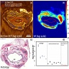
Pahlevaninezhad, Hamid; Lee, Anthony MD; Lam, Stephen; MacAulay, Calum; Lane, Pierre M
Coregistered autofluorescence-optical coherence tomography imaging of human lung sections Journal Article
In: Journal of biomedical optics, vol. 19, no. 3, pp. 036022–036022, 2014.
@article{pahlevaninezhad2014coregistered,

title = {Coregistered autofluorescence-optical coherence tomography imaging of human lung sections},

author = { Hamid Pahlevaninezhad and Anthony MD Lee and Stephen Lam and Calum MacAulay and Pierre M Lane},

url = {https://biophotonics.bccrc.ca/pubs/Pahlevaninezhad-2014a.pdf, Full Text (PDF)

http://www.ncbi.nlm.nih.gov/pubmed/24687614, PubMed},

doi = {10.1117/1.JBO.19.3.036022},

year  = {2014},

date = {2014-01-01},

journal = {Journal of biomedical optics},

volume = {19},

number = {3},

pages = {036022--036022},

publisher = {International Society for Optics and Photonics},

abstract = {Autofluorescence (AF) imaging can provide valuable information about the structural and metabolic state of tissue that can be useful for elucidating physiological and pathological processes. Optical coherence tomography (OCT) provides high resolution detailed information about tissue morphology. We present coregistered AF-OCT imaging of human lung sections. Adjacent hematoxylin and eosin stained histological sections are used to identify tissue structures observed in the OCT images. Segmentation of these structures in the OCT images allowed determination of relative AF intensities of human lung components. Since the AF imaging was performed on tissue sections perpendicular to the airway axis, the results show the AF signal originating from the airway wall components free from the effects of scattering and absorption by overlying layers as is the case during endoscopic imaging. Cartilage and dense connective tissue (DCT) are found to be the dominant fluorescing components with the average cartilage AF intensity about four times greater than that of DCT. The epithelium, lamina propria, and loose connective tissue near basement membrane generate an order of magnitude smaller AF signal than the cartilage fluorescence.},

keywords = {},

pubstate = {published},

tppubtype = {article}

}
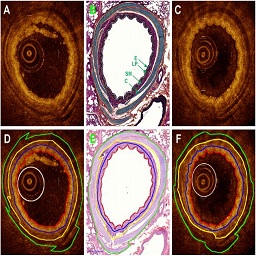
Lee, Anthony MD; Kirby, Miranda; Ohtani, Keishi; Candido, Tara; Shalansky, Rebecca; MacAulay, Calum; English, John; Finley, Richard; Lam, Stephen; Coxson, Harvey O; Lane, Pierre
Validation of airway wall measurements by optical coherence tomography in porcine airways Journal Article
In: PloS one, vol. 9, no. 6, pp. e100145, 2014.
@article{lee2014validation,

title = {Validation of airway wall measurements by optical coherence tomography in porcine airways},

author = { Anthony MD Lee and Miranda Kirby and Keishi Ohtani and Tara Candido and Rebecca Shalansky and Calum MacAulay and John English and Richard Finley and Stephen Lam and Harvey O Coxson and Pierre Lane },

url = {https://biophotonics.bccrc.ca/pubs/Lee-2014a.pdf, Full Text (PDF)

http://www.ncbi.nlm.nih.gov/pubmed/24949633, Pubmed},

doi = {10.1371/journal.pone.0100145},

year  = {2014},

date = {2014-01-01},

journal = {PloS one},

volume = {9},

number = {6},

pages = {e100145},

abstract = {Examining and quantifying changes in airway morphology is critical for studying longitudinal pathogenesis and interventions in diseases such as chronic obstructive pulmonary disease and asthma. Here we present fiber-optic optical coherence tomography (OCT) as a nondestructive technique to precisely and accurately measure the 2-dimensional cross-sectional areas of airway wall substructure divided into the mucosa (WAmuc), submucosa (WAsub), cartilage (WAcart), and the airway total wall area (WAt). Porcine lung airway specimens were dissected from freshly resected lung lobes (N = 10). Three-dimensional OCT imaging using a fiber-optic rotary-pullback probe was performed immediately on airways greater than 0.9 mm in diameter on the fresh airway specimens and subsequently on the same specimens post-formalin-fixation. The fixed specimens were serially sectioned and stained with H&E. OCT images carefully matched to selected sections stained with Movat's pentachrome demonstrated that OCT effectively identifies airway epithelium, lamina propria, and cartilage. Selected H&E sections were digitally scanned and airway total wall areas were measured. Traced measurements of WAmuc, WAsub, WAcart, and WAt from OCT images of fresh specimens by two independent observers found there were no significant differences (p>0.05) between the observer's measurements. The same wall area measurements from OCT images of formalin-fixed specimens found no significant differences for WAsub, WAcart and WAt, and a small but significant difference for WAmuc. Bland-Altman analysis indicated there were negligible biases between the observers for OCT wall area measurements in both fresh and formalin-fixed specimens. Bland-Altman analysis also indicated there was negligible bias between histology and OCT wall area measurements for both fresh and formalin-fixed specimens. We believe this study sets the groundwork for quantitatively monitoring pathogenesis and interventions in the airways using OCT.},

keywords = {},

pubstate = {published},

tppubtype = {article}

}
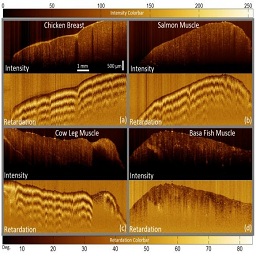
Pahlevaninezhad, Hamid; Lee, Anthony; Cahill, Lucas; Lam, Stephen; MacAulay, Calum; Lane, Pierre
Fiber-Based Polarization Diversity Detection for Polarization-Sensitive Optical Coherence Tomography Journal Article
In: Photonics, vol. 1, no. 4, pp. 283–295, 2014.
@article{pahlevaninezhad2014fiber,

title = {Fiber-Based Polarization Diversity Detection for Polarization-Sensitive Optical Coherence Tomography},

author = { Hamid Pahlevaninezhad and Anthony Lee and Lucas Cahill and Stephen Lam and Calum MacAulay and Pierre Lane},

url = {https://biophotonics.bccrc.ca/pubs/Pahlevaninezhad-2014b.pdf, Full Text (PDF)},

doi = {10.3390/photonics1040283},

year  = {2014},

date = {2014-01-01},

journal = {Photonics},

volume = {1},

number = {4},

pages = {283--295},

publisher = {Multidisciplinary Digital Publishing Institute},

abstract = {We present a new fiber-based polarization diversity detection (PDD) scheme for polarization sensitive optical coherence tomography (PSOCT). This implementation uses a new custom miniaturized polarization-maintaining fiber coupler with single mode (SM) fiber inputs and polarization maintaining (PM) fiber outputs. The SM fiber inputs obviate matching the optical lengths of the two orthogonal OCT polarization channels prior to interference while the PM fiber outputs ensure defined orthogonal axes after interference. Advantages of this detection scheme over those with bulk optics PDD include lower cost, easier miniaturization, and more relaxed alignment and handling issues. We incorporate this PDD scheme into a galvanometer-scanned OCT system to demonstrate system calibration and PSOCT imaging of an achromatic quarter-wave plate, fingernail in vivo, and chicken breast, salmon, cow leg, and basa fish muscle samples ex vivo.},

keywords = {},

pubstate = {published},

tppubtype = {article}

}
Wang, Lu; Lee, Jong Soo; Lane, Pierre; Atkinson, E Neely; Zuluaga, Andres; Follen, Michele; MacAulay, Calum; Cox, Dennis D
A statistical model for removing inter-device differences in spectroscopy Journal Article
In: Optics express, vol. 22, no. 7, pp. 7617–7624, 2014.
@article{wang2014statistical,

title = {A statistical model for removing inter-device differences in spectroscopy},

author = { Lu Wang and Jong Soo Lee and Pierre Lane and E Neely Atkinson and Andres Zuluaga and Michele Follen and Calum MacAulay and Dennis D Cox},

url = {https://biophotonics.bccrc.ca/pubs/Wang-2014.pdf, Full Text (PDF)

http://www.ncbi.nlm.nih.gov/pubmed/24718136, Pubmed},

doi = { 10.1364/OE.22.007617},

year  = {2014},

date = {2014-01-01},

journal = {Optics express},

volume = {22},

number = {7},

pages = {7617--7624},

publisher = {Optical Society of America},

abstract = {We are investigating spectroscopic devices designed to make in vivo cervical tissue measurements to detect pre-cancerous and cancerous lesions. All devices have the same design and ideally should record identical measurements. However, we observed consistent differences among them. An experiment was designed to study the sources of variation in the measurements recorded. Here we present a log additive statistical model that incorporates the sources of variability we identified. Based on this model, we estimated correction factors from the experimental data needed to eliminate the inter-device variability and other sources of variation. These correction factors are intended to improve the accuracy and repeatability of such devices when making future measurements on patient tissue.},

keywords = {},

pubstate = {published},

tppubtype = {article}

}
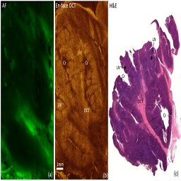
Pahlevaninezhad, Hamid; Lee, Anthony MD; Rosin, Miriam; Sun, Ivan; Zhang, Lewei; Hakimi, Mehrnoush; MacAulay, Calum; Lane, Pierre M
Optical Coherence Tomography and Autofluorescence Imaging of Human Tonsil Journal Article
In: PloS one, vol. 9, no. 12, pp. e115889, 2014.
@article{pahlevaninezhad2014optical,

title = {Optical Coherence Tomography and Autofluorescence Imaging of Human Tonsil},

author = { Hamid Pahlevaninezhad and Anthony MD Lee and Miriam Rosin and Ivan Sun and Lewei Zhang and Mehrnoush Hakimi and Calum MacAulay and Pierre M Lane},

url = {https://biophotonics.bccrc.ca/pubs/Pahlevaninezhad-2014c.pdf, Full Text (PDF)

http://www.ncbi.nlm.nih.gov/pubmed/25542010, Pubmed},

doi = {10.1371/journal.pone.0115889},

year  = {2014},

date = {2014-01-01},

journal = {PloS one},

volume = {9},

number = {12},

pages = {e115889},

publisher = {Public Library of Science},

abstract = {For the first time, we present co-registered autofluorescence imaging and optical coherence tomography (AF/OCT) of excised human palatine tonsils to evaluate the capabilities of OCT to visualize tonsil tissue components. Despite limited penetration depth, OCT can provide detailed structural information about tonsil tissue with much higher resolution than that of computed tomography, magnetic resonance imaging, and Ultrasound. Different tonsil tissue components such as epithelium, dense connective tissue, lymphoid nodules, and crypts can be visualized by OCT. The co-registered AF imaging can provide matching biochemical information. AF/OCT scans may provide a non-invasive tool for detecting tonsillar cancers and for studying the natural history of their development.},

keywords = {},

pubstate = {published},

tppubtype = {article}

}
Lane, Pierre; MacAulay, Calum; Follen, Michele
Endoscopic Confocal Microscopy Book Chapter
In: Zeng, Haishan (Ed.): Chapter 8, pp. 103, Taylor & Francis, 2013, ISBN: 9781420083460.
@inbook{lane20138,

title = {Endoscopic Confocal Microscopy},

author = { Pierre Lane and Calum MacAulay and Michele Follen},

editor = {Haishan Zeng},

url = {https://www.routledge.com/Medical-Equipment-Management/Willson-Ison-Tabakov/p/book/9781420099584},

isbn = {9781420083460},

year  = {2013},

date = {2013-12-09},

urldate = {2013-12-09},

journal = {Diagnostic Endoscopy},

pages = {103},

publisher = {Taylor & Francis},

chapter = {8},

series = {Series in Medical Physics and Biomedical Engineering},

keywords = {},

pubstate = {published},

tppubtype = {inbook}

}
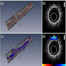
Lee, Anthony MD; Ohtani, Keishi; MacAulay, Calum; McWilliams, Annette; Shaipanich, Tawimas; Yang, Victor XD; Lam, Stephen; Lane, Pierre
In vivo lung microvasculature visualized in three dimensions using fiber-optic color Doppler optical coherence tomography Journal Article
In: Journal of biomedical optics, vol. 18, no. 5, pp. 050501–050501, 2013.
@article{lee2013vivo,

title = {In vivo lung microvasculature visualized in three dimensions using fiber-optic color Doppler optical coherence tomography},

author = { Anthony MD Lee and Keishi Ohtani and Calum MacAulay and Annette McWilliams and Tawimas Shaipanich and Victor XD Yang and Stephen Lam and Pierre Lane},

url = {https://biophotonics.bccrc.ca/pubs/Lee-2013.pdf, Full Text (PDF)

http://www.ncbi.nlm.nih.gov/pubmed/ 23625308, PubMed},

doi = {10.1117/1.JBO.18.5.050501},

year  = {2013},

date = {2013-01-01},

journal = {Journal of biomedical optics},

volume = {18},

number = {5},

pages = {050501--050501},

publisher = {International Society for Optics and Photonics},

abstract = {For the first time, the use of fiber-optic color Doppler optical coherence tomography (CDOCT) to map in vivo the three-dimensional (3-D) vascular network of airway segments in human lungs is demonstrated. Visualizing the 3-D vascular network in the lungs may provide new opportunities for detecting and monitoring lung diseases such as asthma, chronic obstructive pulmonary disease, and lung cancer. Our CDOCT instrument employs a rotary fiber-optic probe that provides simultaneous two-dimensional (2-D) real-time structural optical coherence tomography (OCT) and CDOCT imaging at frame rates up to 12.5 frames per second. Controlled pullback of the probe allows 3-D vascular mapping in airway segments up to 50 mm in length in a single acquisition. We demonstrate the ability of CDOCT to map both small and large vessels. In one example, CDOCT imaging allows assignment of a feature in the structural OCT image as a large (∼1  mm diameter) blood vessel. In a second example, a smaller vessel (∼80  μm diameter) that is indistinguishable in the structural OCT image is fully visualized in 3-D using CDOCT.},

keywords = {},

pubstate = {published},

tppubtype = {article}

}
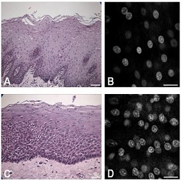
Hallani, S El; Poh, CF; Macaulay, CE; Follen, M; Guillaud, M; Lane, P
Ex vivo confocal imaging with contrast agents for the detection of oral potentially malignant lesions Journal Article
In: Oral oncology, vol. 49, no. 6, pp. 582–590, 2013.
@article{el2013ex,

title = {Ex vivo confocal imaging with contrast agents for the detection of oral potentially malignant lesions},

author = { S El Hallani and CF Poh and CE Macaulay and M Follen and M Guillaud and P Lane},

url = {https://biophotonics.bccrc.ca/pubs/Hallani-2013.pdf, Full Text (PDF)

http://www.ncbi.nlm.nih.gov/pubmed/23415144, PubMed},

doi = {10.1016/j.oraloncology.2013.01.009},

year  = {2013},

date = {2013-01-01},

journal = {Oral oncology},

volume = {49},

number = {6},

pages = {582--590},

publisher = {Elsevier},

abstract = {Objectives


We investigated the potential use of real-time confocal microscpy in the non-invasive detection of occult oral potentially malignant lesions. Our objectives were to select the best fluorescence contrast agent for cellular morphology enhancement, to build an atlas of confocal microscopic images of normal human oral mucosa, and to determine the accuracy of confocal microscopy to recognise oral high-grade dysplasia lesions on live human tissue.


Materials and Methods


Five clinically used fluorescent contrast agents were tested in vitro on cultured human cells and validated ex vivo on human oral mucosa. Images acquired ex vivo from normal and diseased human oral biopsies with bench-top fluorescent confocal microscope were compared to conventional histology. Image analyzer software was used as an adjunct tool to objectively compare high-grade dysplasia versus low-grade dysplasia and normal epithelium.


Results


Acriflavine Hydrochloride provided the best cellular contrast by preferentially staining the nuclei of the epithelium. Using topical application of Acriflavine Hydrochloride followed by confocal microscopy, we could define morphological characteristics of each cellular layer of the normal human oral mucosa, building an atlas of histology-like images. Applying this technique to diseased oral tissue specimen, we were also able to accurately diagnose the presence of high-grade dysplasia through the increased cellularity and changes in nuclear morphological features. Objective measurement of cellular density by quantitative image analysis was a strong discriminant to differentiate between high-grade dysplasia and low-grade dysplasia lesions.


Conclusions


Pending clinical investigation, real-time confocal microscopy may become a useful adjunct to detect precancerous lesions that are at high risk of cancer progression, direct biopsy and delineate excision margins.},

keywords = {},

pubstate = {published},

tppubtype = {article}

}
We investigated the potential use of real-time confocal microscpy in the non-invasive detection of occult oral potentially malignant lesions. Our objectives were to select the best fluorescence contrast agent for cellular morphology enhancement, to build an atlas of confocal microscopic images of normal human oral mucosa, and to determine the accuracy of confocal microscopy to recognise oral high-grade dysplasia lesions on live human tissue.
Materials and Methods
Five clinically used fluorescent contrast agents were tested in vitro on cultured human cells and validated ex vivo on human oral mucosa. Images acquired ex vivo from normal and diseased human oral biopsies with bench-top fluorescent confocal microscope were compared to conventional histology. Image analyzer software was used as an adjunct tool to objectively compare high-grade dysplasia versus low-grade dysplasia and normal epithelium.
Results
Acriflavine Hydrochloride provided the best cellular contrast by preferentially staining the nuclei of the epithelium. Using topical application of Acriflavine Hydrochloride followed by confocal microscopy, we could define morphological characteristics of each cellular layer of the normal human oral mucosa, building an atlas of histology-like images. Applying this technique to diseased oral tissue specimen, we were also able to accurately diagnose the presence of high-grade dysplasia through the increased cellularity and changes in nuclear morphological features. Objective measurement of cellular density by quantitative image analysis was a strong discriminant to differentiate between high-grade dysplasia and low-grade dysplasia lesions.
Conclusions
Pending clinical investigation, real-time confocal microscopy may become a useful adjunct to detect precancerous lesions that are at high risk of cancer progression, direct biopsy and delineate excision margins.
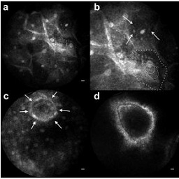
Contaldo, Maria; Poh, Catherine F; Guillaud, Martial; Lucchese, Alberta; Rullo, Rosario; Lam, Sylvia; Serpico, Rosario; MacAulay, Calum E; Lane, Pierre M
In: Oral surgery, oral medicine, oral pathology and oral radiology, vol. 116, no. 6, pp. 752–758, 2013.
@article{contaldo2013oral,

title = {Oral mucosa optical biopsy by a novel handheld fluorescent confocal microscope specifically developed: technologic improvements and future prospects},

author = { Maria Contaldo and Catherine F Poh and Martial Guillaud and Alberta Lucchese and Rosario Rullo and Sylvia Lam and Rosario Serpico and Calum E MacAulay and Pierre M Lane},

url = {https://biophotonics.bccrc.ca/pubs/Contaldo-2013.pdf, Full Text (PDF)

http://www.ncbi.nlm.nih.gov/pubmed/24237726, Pubmed},

doi = {10.1016/j.oooo.2013.09.006},

year  = {2013},

date = {2013-01-01},

journal = {Oral surgery, oral medicine, oral pathology and oral radiology},

volume = {116},

number = {6},

pages = {752--758},

publisher = {Elsevier},

abstract = {Objective


This pilot study evaluated the baseline effectiveness of a novel handheld fluorescent confocal microscope (FCM) specifically developed for oral mucosa imaging and compared the results with the literature.


Study Design


Four different oral sites (covering the mucosa of the lip and of the ventral tongue, the masticatory mucosa of the gingiva, and the specialized mucosa of the dorsal tongue) in 6 healthy nonsmokers were imaged by an FCM made up of a confocal fiberoptic probe ergonomically designed for in vivo oral examination, using light at the wavelength of 457 nm able to excite the fluorophore acriflavine hydrochloride, topically administered. In total, 24 mucosal areas were examined.


Results


The FCM was able to distinctly define epithelial cells, bacterial plaque, and inflammatory cells and to image submucosal structures by detecting their intrinsic fluorescence.


Conclusions


When compared with other devices, this FCM allowed the user to image each oral site at higher magnification, thus resulting in a clearer view.},

keywords = {},

pubstate = {published},

tppubtype = {article}

}
This pilot study evaluated the baseline effectiveness of a novel handheld fluorescent confocal microscope (FCM) specifically developed for oral mucosa imaging and compared the results with the literature.
Study Design
Four different oral sites (covering the mucosa of the lip and of the ventral tongue, the masticatory mucosa of the gingiva, and the specialized mucosa of the dorsal tongue) in 6 healthy nonsmokers were imaged by an FCM made up of a confocal fiberoptic probe ergonomically designed for in vivo oral examination, using light at the wavelength of 457 nm able to excite the fluorophore acriflavine hydrochloride, topically administered. In total, 24 mucosal areas were examined.
Results
The FCM was able to distinctly define epithelial cells, bacterial plaque, and inflammatory cells and to image submucosal structures by detecting their intrinsic fluorescence.
Conclusions
When compared with other devices, this FCM allowed the user to image each oral site at higher magnification, thus resulting in a clearer view.
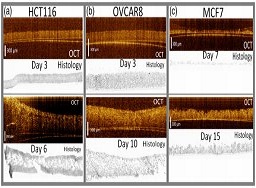
Pahlevaninezhad, Hamid; Cecic, Ivana; Lee, Anthony MD; Kyle, Alastair H; Lam, Stephen; MacAulay, Calum; Lane, Pierre M
In: Journal of biomedical optics, vol. 18, no. 10, pp. 106007–106007, 2013.
@article{pahlevaninezhad2013multimodal,

title = {Multimodal tissue imaging: using coregistered optical tomography data to estimate tissue autofluorescence intensity change due to scattering and absorption by neoplastic epithelial cells},

author = { Hamid Pahlevaninezhad and Ivana Cecic and Anthony MD Lee and Alastair H Kyle and Stephen Lam and Calum MacAulay and Pierre M Lane},

url = {https://biophotonics.bccrc.ca/pubs/Pahlevaninezhad-2013.pdf, Full Text (PDF)

http://www.ncbi.nlm.nih.gov/pubmed/24108573, Pubmed},

doi = {10.1117/1.JBO.18.10.106007},

year  = {2013},

date = {2013-01-01},

journal = {Journal of biomedical optics},

volume = {18},

number = {10},

pages = {106007--106007},

publisher = {International Society for Optics and Photonics},

abstract = {Autofluorescence (AF) imaging provides valuable information about the structural and chemical states of tissue that can be used for early cancer detection. Optical scattering and absorption of excitation and emission light by the epithelium can significantly affect observed tissue AF intensity. Determining the effect of epithelial attenuation on the AF intensity could lead to a more accurate interpretation of AF intensity. We propose to use optical coherence tomography coregistered with AF imaging to characterize the AF attenuation due to the epithelium. We present imaging results from three vital tissue models, each consisting of a three-dimensional tissue culture grown from one of three epithelial cell lines (HCT116, OVCAR8, and MCF7) and immobilized on a fluorescence substrate. The AF loss profiles in the tissue layer show two different regimes, each approximately linearly decreasing with thickness. For thin cell cultures (<300 μm), the AF signal changes as AF(t)/AF(0)=1-1.3t (t is the thickness in millimeter). For thick cell cultures (>400 μm), the AF loss profiles have different intercepts but similar slopes. The data presented here can be used to estimate AF loss due to a change in the epithelial layer thickness and potentially to reduce AF bronchoscopy false positives due to inflammation and non-neoplastic epithelial thickening.},

keywords = {},

pubstate = {published},

tppubtype = {article}

}
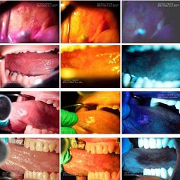
Lane, Pierre; Follen, Michele; MacAulay, Calum
In: Gender medicine, vol. 9, no. 1, pp. S25–S35, 2012.
@article{lane2012has,

title = {Has fluorescence spectroscopy come of age? A case series of oral precancers and cancers using white light, fluorescent light at 405 nm, and reflected light at 545 nm using the Trimira Identafi 3000},

author = { Pierre Lane and Michele Follen and Calum MacAulay},

url = {https://biophotonics.bccrc.ca/pubs/Lane-2012.pdf, Full Text (PDF)

http://www.ncbi.nlm.nih.gov/pubmed/22340638, PubMed},

doi = {10.1016/j.genm.2011.09.031},

year  = {2012},

date = {2012-01-01},

journal = {Gender medicine},

volume = {9},

number = {1},

pages = {S25--S35},

publisher = {Elsevier},

abstract = {Objectives


The objective of this study was to present a case series of oral precancers and cancers that have been photographed during larger ongoing clinical trials.


Methods


Over 300 patients were measured at 2 clinical sites that are comprehensive cancer centers and a faculty practice associated with a major dental school. Each site is conducting independent research on the sensitivity and specificity of several optical technologies for the diagnosis of oral neoplasia. The cases presented in this case series were taken from the larger database of images from the clinical trials using the aforementioned device. Optical spectroscopy was performed and biopsies obtained from all sites measured, representing abnormal and normal areas on comprehensive white light examination and after use of the fluorescence and reflectance spectroscopy device. The gold standard of test accuracy was the histologic report of biopsies read by the study histopathologists at each of the 3 study sites.


Results


Comprehensive white light examination showed some lesions; however, the addition of a fluorescence image and a selected reflectance wavelength was helpful in identifying other characteristics of the lesions. The addition of the violet light-induced fluorescence excited at 405 nm provided an additional view of both the stromal neovasculature of the lesions and the stromal changes associated with lesion growth that were biologically indicative of stromal breakdown. The addition of 545 nm green–amber light reflectance increased the view of the keratinized image and allowed the abnormal surface vasculature to be more prominent.


Conclusions


Optical spectroscopy is a promising technology for the diagnosis of oral neoplasia. The conclusion of several ongoing clinical trials and an eventual randomized Phase III clinical trial will provide definitive findings that sensitivity is or is not increased over comprehensive white light examination.},

keywords = {},

pubstate = {published},

tppubtype = {article}

}
The objective of this study was to present a case series of oral precancers and cancers that have been photographed during larger ongoing clinical trials.
Methods
Over 300 patients were measured at 2 clinical sites that are comprehensive cancer centers and a faculty practice associated with a major dental school. Each site is conducting independent research on the sensitivity and specificity of several optical technologies for the diagnosis of oral neoplasia. The cases presented in this case series were taken from the larger database of images from the clinical trials using the aforementioned device. Optical spectroscopy was performed and biopsies obtained from all sites measured, representing abnormal and normal areas on comprehensive white light examination and after use of the fluorescence and reflectance spectroscopy device. The gold standard of test accuracy was the histologic report of biopsies read by the study histopathologists at each of the 3 study sites.
Results
Comprehensive white light examination showed some lesions; however, the addition of a fluorescence image and a selected reflectance wavelength was helpful in identifying other characteristics of the lesions. The addition of the violet light-induced fluorescence excited at 405 nm provided an additional view of both the stromal neovasculature of the lesions and the stromal changes associated with lesion growth that were biologically indicative of stromal breakdown. The addition of 545 nm green–amber light reflectance increased the view of the keratinized image and allowed the abnormal surface vasculature to be more prominent.
Conclusions
Optical spectroscopy is a promising technology for the diagnosis of oral neoplasia. The conclusion of several ongoing clinical trials and an eventual randomized Phase III clinical trial will provide definitive findings that sensitivity is or is not increased over comprehensive white light examination.
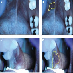
Lane, Pierre; Lam, Sylvia; Follen, Michele; MacAulay, Calum
In: Gender medicine, vol. 9, no. 1, pp. S78–S82, 2012.
@article{lane2012oral,

title = {Oral fluorescence imaging using 405-nm excitation, aiding the discrimination of cancers and precancers by identifying changes in collagen and elastic breakdown and neovascularization in the underlying stroma},

author = { Pierre Lane and Sylvia Lam and Michele Follen and Calum MacAulay},

url = {https://biophotonics.bccrc.ca/pubs/Lane-2012a.pdf, Full Text (PDF)

http://www.ncbi.nlm.nih.gov/pubmed/22340643, PubMed},

doi = {10.1016/j.genm.2011.11.006},

year  = {2012},

date = {2012-01-01},

journal = {Gender medicine},

volume = {9},

number = {1},

pages = {S78--S82},

publisher = {Elsevier},

abstract = {Background


Optical spectroscopy and imaging devices are being developed and tested for the screening and diagnosis of cancer and precancer in multiple organ sites.


Objective


The aim of the study reported here is to optimize the capability of an optical imaging device to discriminate precancerous tissue from other lesions by identifying ideal excitation wavelengths.


Methods


The studies reported here used a prototype of a direct fluorescence imaging device that uses 405-nm illumination to excite tissue.


Results


There is ample evidence in the literature that 405 nm can distinguish oral cancers from normal tissue. Higher wavelengths may be necessary to differentiate potential confounding lesions, such as abrasions, burns, viral infections, inflammation, and gingivitis.


Conclusions


Imaging at 405 nm could help doctors detect precancerous and cancerous oral lesions. Such imaging could be used by dentists, family practitioners, otorhinolaryngologists, general surgeons, obstetrician gynecologists, and internists, and could greatly increase the number of patients who have lesions detected in the precancerous phase.},

keywords = {},

pubstate = {published},

tppubtype = {article}

}
Optical spectroscopy and imaging devices are being developed and tested for the screening and diagnosis of cancer and precancer in multiple organ sites.
Objective
The aim of the study reported here is to optimize the capability of an optical imaging device to discriminate precancerous tissue from other lesions by identifying ideal excitation wavelengths.
Methods
The studies reported here used a prototype of a direct fluorescence imaging device that uses 405-nm illumination to excite tissue.
Results
There is ample evidence in the literature that 405 nm can distinguish oral cancers from normal tissue. Higher wavelengths may be necessary to differentiate potential confounding lesions, such as abrasions, burns, viral infections, inflammation, and gingivitis.
Conclusions
Imaging at 405 nm could help doctors detect precancerous and cancerous oral lesions. Such imaging could be used by dentists, family practitioners, otorhinolaryngologists, general surgeons, obstetrician gynecologists, and internists, and could greatly increase the number of patients who have lesions detected in the precancerous phase.
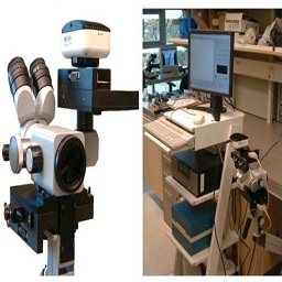
Buys, Timon PH; Cantor, Scott B; Guillaud, Martial; Adler-Storthz, Karen; Cox, Dennis D; Okolo, Clement; Arulogon, Oyedunni; Oladepo, Oladimeji; Basen-Engquist, Karen; Shinn, Eileen; Yamal, José-Miguel; Beck, J. Robert; Scheurer, Michael E.; van Niekerk, Dirk; Malpica, Anais; Matisic, Jasenka; Staerkel, Gregg; Atkinson, Edward Neely; Bidaut, Luc; Lane, Pierre; Benedet, J. Lou; Miller, Dianne; Ehlen, Tom; Price, Roderick; Adewole, Isaac F.; MacAulay, Calum; Follen, Michele
Optical technologies and molecular imaging for cervical neoplasia: a program project update Journal Article
In: Gender medicine, vol. 9, no. 1, pp. S7–S24, 2012.
@article{buys2012optical,

title = {Optical technologies and molecular imaging for cervical neoplasia: a program project update},

author = { Timon PH Buys and Scott B Cantor and Martial Guillaud and Karen Adler-Storthz and Dennis D Cox and Clement Okolo and Oyedunni Arulogon and Oladimeji Oladepo and Karen Basen-Engquist and Eileen Shinn and José-Miguel Yamal and J. Robert Beck and Michael E. Scheurer and Dirk van Niekerk and Anais Malpica and Jasenka Matisic and Gregg Staerkel and Edward Neely Atkinson and Luc Bidaut and Pierre Lane and J. Lou Benedet and Dianne Miller and Tom Ehlen and Roderick Price and Isaac F. Adewole and Calum MacAulay and Michele Follen },

url = {https://biophotonics.bccrc.ca/pubs/Buys-2012.pdf, Full Text (PDF)

http://www.ncbi.nlm.nih.gov/pubmed/21944317, PubMed},

doi = {10.1016/j.genm.2011.08.002},

year  = {2012},

date = {2012-01-01},

journal = {Gender medicine},

volume = {9},

number = {1},

pages = {S7--S24},

publisher = {Elsevier},

abstract = {There is an urgent global need for effective and affordable approaches to cervical cancer screening and diagnosis. In developing nations, cervical malignancies remain the leading cause of cancer-related deaths in women. This reality may be difficult to accept given that these deaths are largely preventable; where cervical screening programs have been implemented, cervical cancer-related deaths have decreased dramatically. In developed countries, the challenges of cervical disease stem from high costs and overtreatment. The National Cancer Institute-funded Program Project is evaluating the applicability of optical technologies in cervical cancer. The mandate of the project is to create tools for disease detection and diagnosis that are inexpensive, require minimal expertise, are more accurate than existing modalities, and can be feasibly implemented in a variety of clinical settings. This article presents the status and long-term goals of the project.},

keywords = {},

pubstate = {published},

tppubtype = {article}

}


