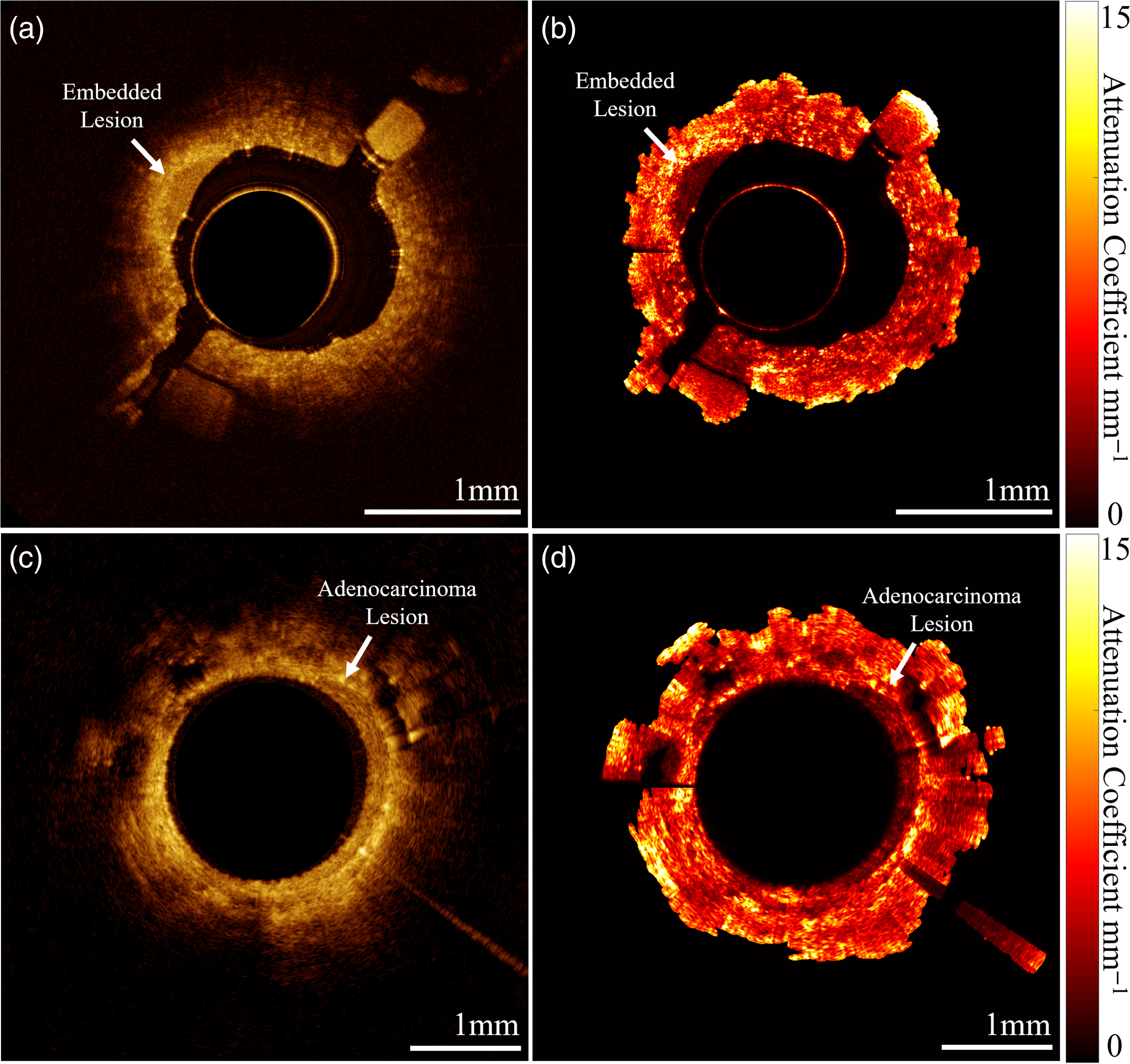


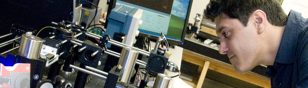


Eric Brace, a PhD student in our group has a new publication in Optica’s Biomedical Optics Express.
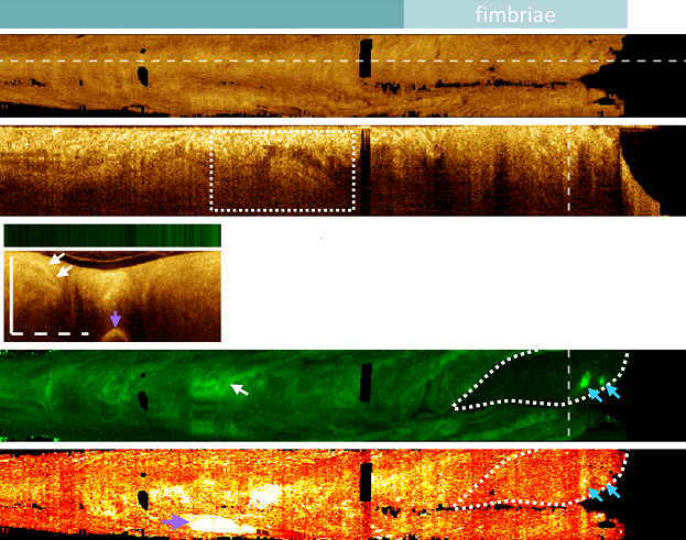
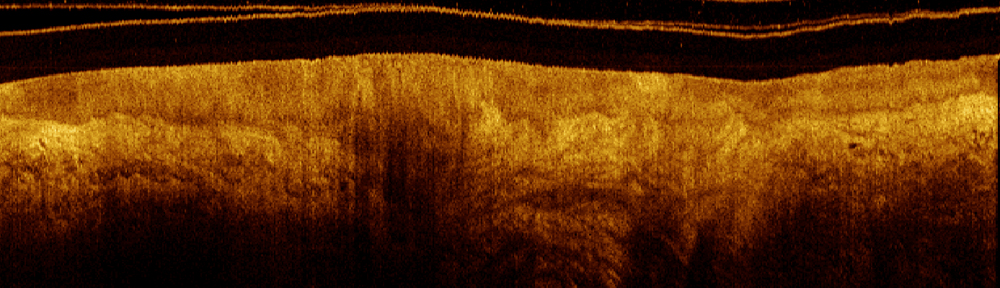
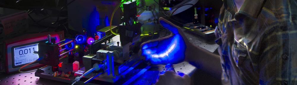
Blue-light autofluorescence imaging captures a snapshot of tissue biochemistry without using dyes or contrast agents. Because healthy and abnormal tissues produce different fluorescence patterns, this approach can be used to distinguish cancerous or precancerous lesions.
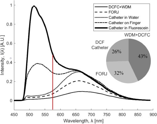
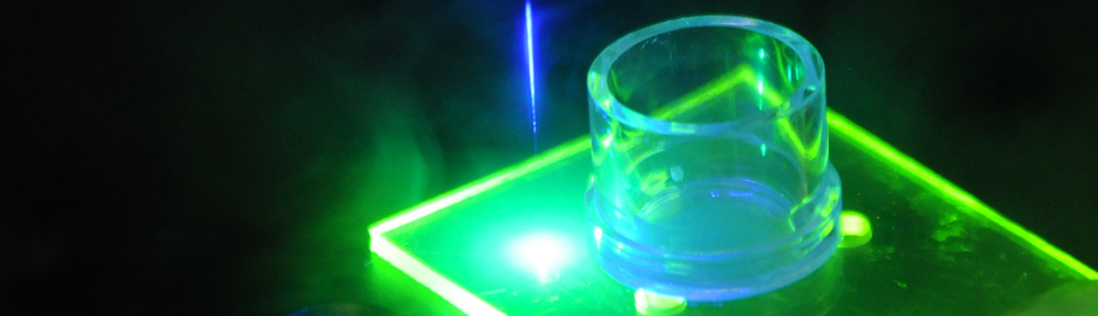
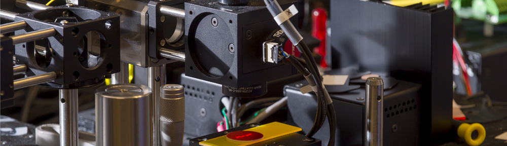
However, the amount of natural fluorescence from tissue is very faint. Additionally, parts of the imaging system (like plastics) can fluoresce, introducing unwanted background light that makes the fluorescence from tissue harder to detect.
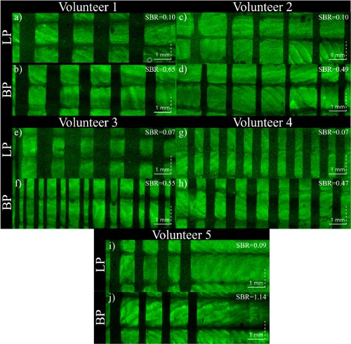
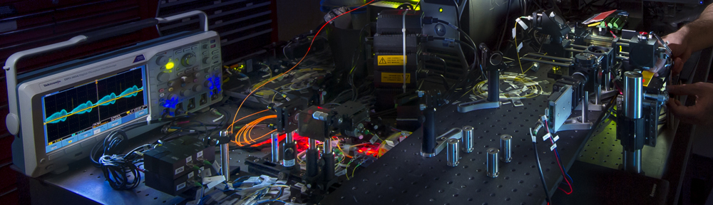
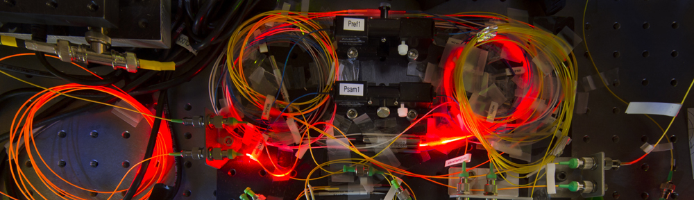
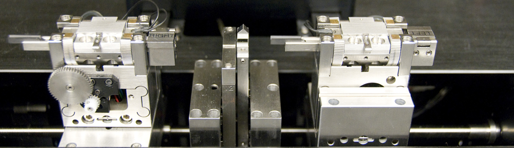
This work characterized the fluorescence of each component in a rotary pullback catheter OCT-AFI system to identify sources of unwanted background fluorescence. From this analysis, modifications were made to the imaging system including a new filtering approach. The result was a nearly eightfold improvement in the system’s signal-to-background ratio.
This improvement makes it possible to identify subtler features in autofluorescence imaging, which we hope will support detecting earlier or smaller lesions.