


Congratulations to Kimiya Mousavi on successfully defending her MASc thesis!
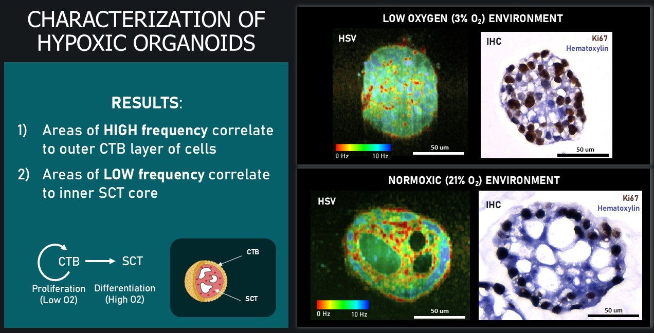
Kimiya’s work ‘Label-Free Vital Imaging Of Trophoblast Organoids With Dynamic Optical Coherence Microscopy’ focused on developing imaging techniques to study placental models. Using an OCM system, Kimiya collected high-resolution videos of these organoids and applied three algorithms which generate contrast based on the metabolic activity (motion) within cells.
Kimiya developed quantitative methods to distinguish trophoblast organoids grown under low-oxygen conditions (which mimics complications like preeclampsia) and those grown in normal oxygen levels. This is an important step toward better understanding how the placenta develops under different conditions.
A key advantage of this technique is that it allows researchers to monitor living tissues over time without damaging them. Because it avoids the need for sectioning, staining, or dyes which disrupt biologic systems, it preserves the biology of the system and opens the door to longitudinal studies which could strengthen our understanding of placental health and disease.