


Jeanie, a PhD student in our lab has a new publication in MDPI Cancers.
Oral cancers are associated with high mortality in advanced stages. Early diagnosis is associated with better patient outcomes, but this is challenging to achieve as benign lesions look similar to lesions of concern, and multiple biopsies may be required to ensure the most pathologic tissue is sampled.
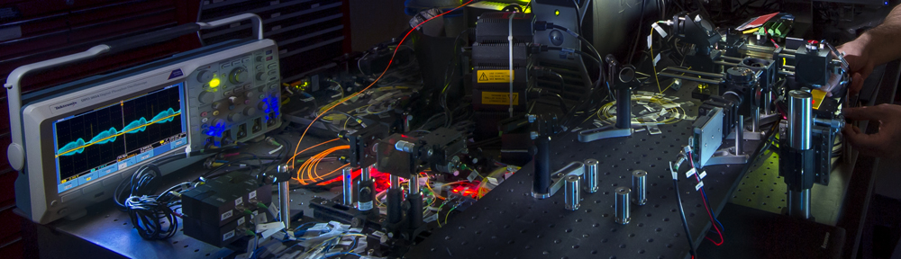
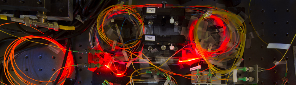
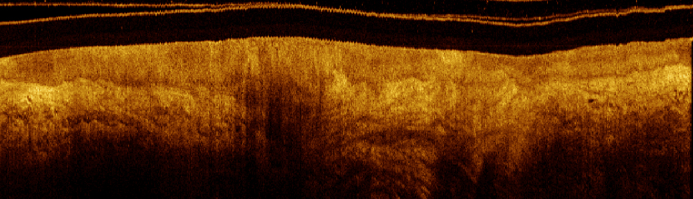
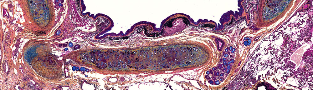
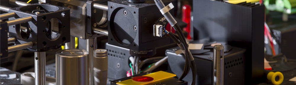
Optical coherence tomography is a noninvasive imaging technique that provides three-dimensional visualization of subsurface tissue structures. We have previously developed an OCT endoscope which can reach most sites in the oral cavity – but assessing large three-dimensional volumes for the features most relevant to oral cancer is challenging.
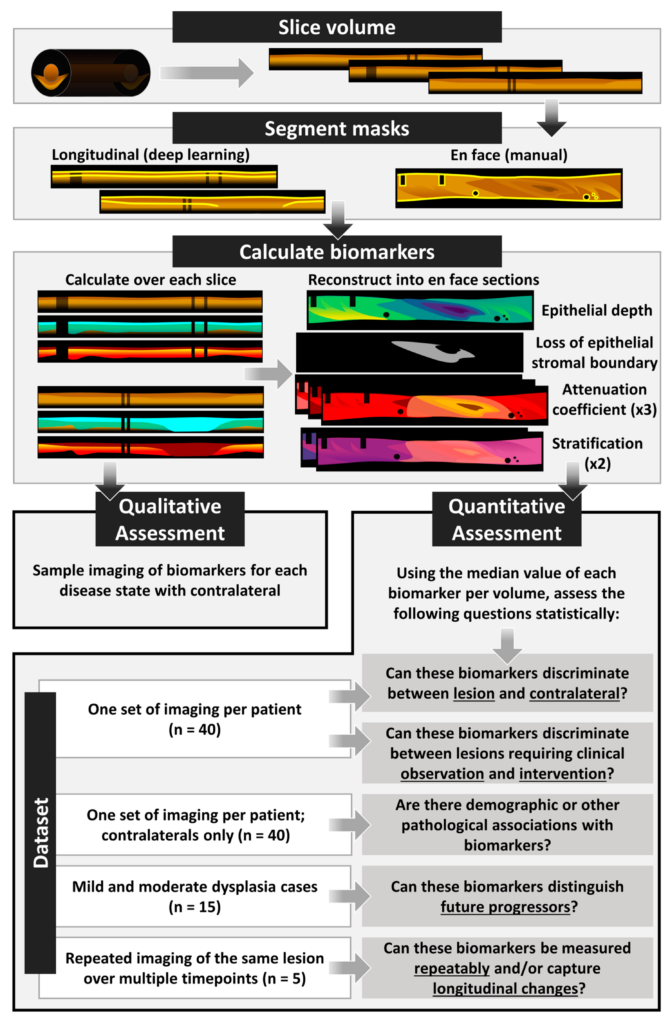
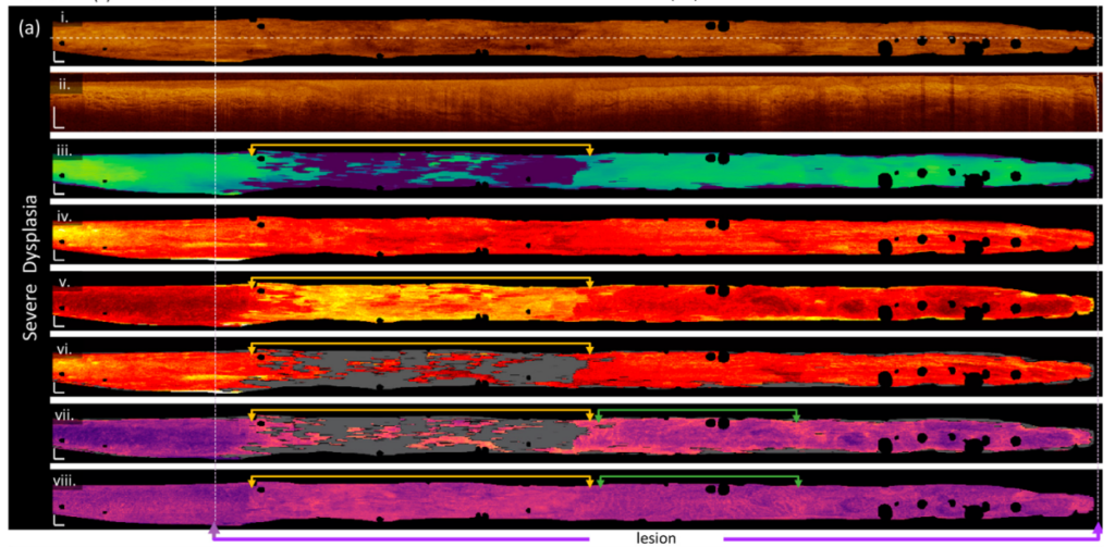
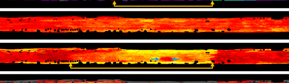
This work leverages the work of Chloe, a recent Masters student in our lab, who developed a deep learning segmentation tool to rapidly detect the tissue surface and bottom of the epithelium in oral OCT. From these segmentations, we measure seven imaging biomarkers and assess their utility in distinguishing oral pre-cancers and cancers.